Review
. 2018 Aug;9(4):463-476.
doi: 10.1007/s13244-018-0605-6. Epub 2018 Apr 11.

Imaging of urgencies and emergencies in the lung cancer patient

Affiliations
Review

Imaging of urgencies and emergencies in the lung cancer patient

Bruno De Potter et al. Insights Imaging. 2018 Aug.

Abstract

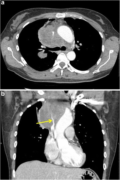
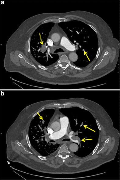
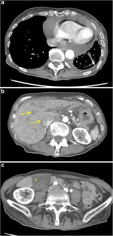
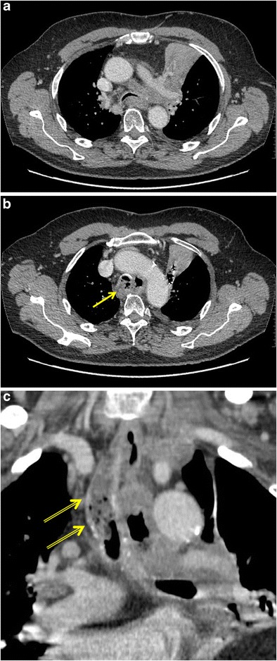
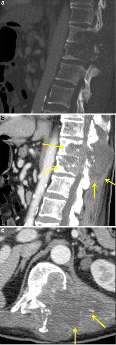
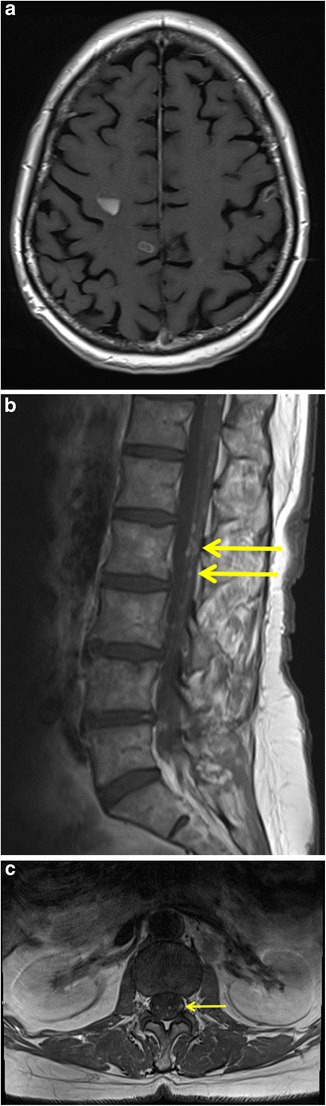
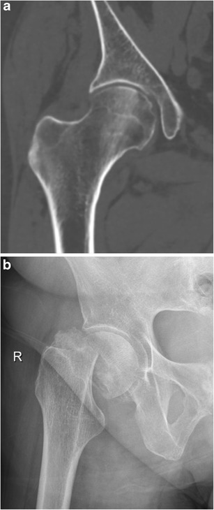
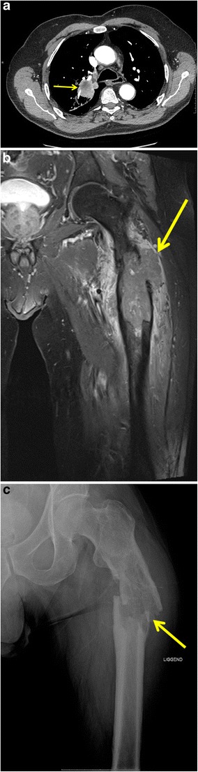
Lung cancer patients often experience potentially life-threatening medical urgencies and emergencies, which may be a direct or indirect result of the underlying malignancy. This pictorial review addresses the most common thoracic, neurological and musculoskeletal medical emergencies in lung cancer patients, including superior vena cava syndrome, pulmonary embolism, spontaneous pneumothorax, cardiac tamponade, massive haemoptysis, central airway obstruction, oesophagorespiratory fistula, malignant spinal cord compression, carcinomatous meningitis, cerebral herniation and pathological fracture. Emphasis is placed on imaging findings, the role of different imaging techniques and a brief discussion of epidemiology, pathophysiology and therapeutic options. Since early diagnosis is important for adequate patient management and prognosis, radiologists have a crucial role in recognising and communicating these urgencies and emergencies.

Teaching points: • Multiplanar multidetector computed tomography is the imaging examination of choice for thoracic urgencies and emergencies. • Magnetic resonance imaging is the imaging modality of choice for investigating central nervous system emergencies. • Urgencies and emergencies can be the initial manifestation of lung cancer. • Radiologists have a crucial role in recognising and in communicating these urgencies/emergencies.

Keywords: Computed tomography; Emergencies; Lung cancer; Magnetic resonance imaging; Radiography.


Conflict of interest statement

Conflicts of interest

All the authors declare that they have no conflict of interest.

Ethical adherence

The material in the manuscript has been acquired according to modern ethical standards.

Figures

Fig. 1
Fig. 1
Superior vena cava syndrome (SVCS) as the initial presentation of a stage IIIB small cell lung cancer in a 48-year-old woman. a Contrast-enhanced axial CT image in mediastinal window setting depicts a large mass located in the visceral mediastinal compartment with encasement of mediastinum and in particular the SVC. b CT image reconstruction in the coronal plane shows the large soft tissue mass and better depicts the prominent narrowing of the SVC (yellow arrow)
Fig. 2
Fig. 2
Pulmonary embolism in a 73-year-old man with known stage IV non-small cell lung cancer. Axial reconstructions of a CT pulmonary angiography study at the carinal (a) and infracarinal (b) level showing a hilar mass (double yellow arrow) in the right upper lobe (a) and large emboli involving the left main pulmonary arteries as well as bilateral subsegmental emboli (a, b) (yellow arrows)
Fig. 3
Fig. 3
Spontaneous pneumothorax as the first sign of a primary lung carcinoma in an 87-year-old woman who presented at the emergency department with thoracic pain and severe respiratory distress. a Supine anteroposterior chest radiograph clearly depicts a right-sided pneumothorax with complete collapse of the right lung. The visceral pleural edge is observed clearly as a very thin, sharp line (yellow arrows) with the absence of vascular marking beyond the pleural line. b Axial contrast-enhanced chest CT in lung window setting shows a large thin-walled cystic lesion (asterisk) with peripheral solid nodular component (yellow arrow) in the right upper lobe as the underlying cause of the pneumothorax. Also, note the extensive subcutaneous emphysema in the right chest wall. Histopathological diagnosis of adenocarcinoma (type “lung cancer associated with cystic airspaces”) was made after lobectomy
Fig. 4
Fig. 4
A 73-year-old woman with a known stage IV non-small cell lung cancer presented during follow-up with symptoms of chest pain, increasing dyspnea and fatigue. a Contrast-enhanced axial CT image in mediastinal window setting depicts a massive pericardial effusion without prominent pericardial thickening, nodularities or enhancement. b, c Axial CT images at the level of the upper abdomen show an enlargement of the inferior vena cava with contrast reflux (double yellow arrow), periportal oedema (yellow arrow) and gallbladder oedema (yellow asterisk) as signs of right heart failure. Findings are compatible with cardiac tamponade. The woman was referred for urgent cardiac work-up and drainage of the cardiac fluid
Fig. 5
Fig. 5
Massive haemoptysis and fatal air embolism in a 68-year-old woman with a cavernous lung lesion. a Anteroposterior chest radiograph, performed for inflammatory changes on blood tests and tachypnea, shows a giant cavitated consolidation in the right upper lobe. b Axial contrast-enhanced chest CT image confirmed a large ill-defined mass with central cavitation (yellow asterisks). Immediately following the CT, the patient developed massive haemoptysis in the radiology department necessitating intubation and ventilation. c Axial-contrast enhanced CT image of the chest after haemodynamic stabilisation shows a large amount of high-density fluid, compatible with blood (yellow arrow), in the cavern and the tracheobronchial tree. In addition, there was massive subcutaneous emphysema, a large amount of air in the vascular system and heart. Upon completion of the scan a fatal cardiac arrest ensued. Histopathology of the post-mortem examination confirmed a stage IV non-small cell lung cancer (poorly differentiated carcinoma)
Fig. 6
Fig. 6
A 64-year-old woman was referred to the pulmonologist for persistent cough and increasing dyspnea. a Erect postero-anterior chest radiograph at the time of presentation shows an enlargement of the left hilum, air trapping in the left hemithorax (suggestive of partial bronchial obstruction) and a left-sided pleural effusion. b Axial contrast-enhanced chest CT in mediastinal window 1 day after the chest radiograph shows a left hilar mass with infracarinal extension, c partially obstructing the left main bronchus (yellow arrow). Although there is only a short time frame between the radiograph and the CT, there is marked volume loss of the left lung and mediastinal shift caused by retro-obstructive atelectasis. Diagnosis of stage IV non-small cell lung cancer was made. Four days after the CT examination, the patient experienced an acute episode of severe dyspnea and stridor for which she was referred to the emergency department. d Erect postero-anterior chest radiograph at admission shows a white lung on the left with massive mediastinal and cardiac shift, caused by complete obstruction of the left main bronchus. No air can be delineated in the left main bronchus. This was confirmed by bronchoscopy
Fig. 7
Fig. 7
Tracheo-oesophageal fistula in a 60-year-old man with known stage IV non-small cell lung cancer who presented with a fever and had complaints of dysphagia. a Axial contrast-enhanced CT in mediastinal window setting shows a large left hilar mass encasing the left pulmonary artery with associated retro-obstructive atelectasis of the left upper lobe. Note mediastinal invasion extending to the trachea and oesophagus (yellow arrow). b Axial CT image at the level of the aortic arch reveals an extraluminal gas collection adjacent to both oesophagus and trachea. Both the walls of the oesophagus and trachea are thickened at this level with blurring and fatty infiltration of the surrounding fat planes. c Reformatted image in the coronal plane nicely depicts the presence of fluid, debris and air-bubbles in the trachea (double yellow arrows). The combination of findings is indicative of a tracheo-oesophageal fistula
Fig. 8
Fig. 8
Malignant spinal cord compression of the thoracic spine in a 56-year-old man with stage IV non-small cell lung cancer. Sagittal enhanced CT image in bone (a) and soft-tissue (b) window setting at the thoracolumbar level shows confluent lytic bone lesions in the T11 and T12 vertebrae with disruption of the posterior wall and extending into the posterior elements. Also, note the large soft-tissue mass (yellow arrows) extending in the spinal canal resulting in a compression of the thoracic spinal cord. Axial enhanced CT image in soft tissue window (c) clearly shows the extension of the soft tissue mass in the spinal canal, while the spinal cord is no longer discernible
Fig. 9
Fig. 9
Carcinomatous meningitis in a 67-year-old woman with stage IV non-small cell lung cancer. Coronal (a) and sagittal (b, c) T1-weighted MR images after intravenous gadolinium contrast administration demonstrate diffuse, nodular leptomeningeal enhancement involving both cerebral hemispheres, but more markedly along the tentorium and into the subarachnoid spaces between the cerebellar folia (yellow arrows)
Fig. 10
Fig. 10
Intradural extramedullary metastases in a 54-year-old woman with non-small cell lung cancer. Axial T1-weighted image of the brain after intravenous gadolinium contrast administration (a) shows presence of multiple ring-enhancing parenchymal lesions consistent with cerebral metastases. Sagittal (b) and axial (c) T1-weighted MR images of the lumbar spine (after intravenous gadolinium contrast administration) demonstrate multiple enhancing tumour nodules (yellow arrows) along the lumbar spinal cord and cauda equina indicating leptomeningeal metastatic disease
Fig. 11
Fig. 11
Cerebral herniation secondary to a cerebellar brain metastasis in a 51-year-old woman non-small lung cancer patient with a history of pancranial radiation therapy for brain metastases. Axial FLAIR (a) and axial T1-weighted image after intravenous gadolinium contrast administration (b) show a peripheral enhancing mass in the left cerebellar hemisphere with peritumoural oedema resulting in a midline shift and compression of the fourth ventricle. Sagittal T1-weighted contrast-enhanced image (c) demonstrates herniation of the left cerebellar tonsil trough the foramen magnum (yellow arrow). Coronal T1-weighted contrast-enhanced images shows an ascending transtentorial herniation with displacement of cerebellar tissue through the tentorial notch (double yellow arrow)
Fig. 12
Fig. 12
Subcapital insufficiency fracture of the right femur in a 77-year-old woman with stage IIIB non-small cell lung cancer. The patient developed extensive radiation pneumonitis, which was treated with high-dose glucocorticoids for 1 year, probably causing a greater rate of bone loss with subsequent insufficiency fracture. a Coronal non-enhanced CT image in bone window, performed 1 month prior to the fracture, shows no signs of metastatic bone involvement in the right hip. b Supine X-ray of the right hip shows a subcapital fracture of the right femur without associated osteolytic bone destruction. Absence of malignancy was confirmed on histopathological examination of the femoral head after total hip replacement
Fig. 13
Fig. 13
Pathological femoral shaft fracture in a 63-year-old man with a stage IV bronchogenic carcinoma. a Axial chest CT image in mediastinal window setting shows a large tumour in the right upper lobe (yellow arrow). b Coronal FS T1-weighted MR image after gadolinium contrast administration depicts a mass in the proximal left femoral shaft with extra-osseous soft tissue component (yellow arrow) and significant surrounding soft-tissue oedema. The patient presented to the emergency department 1 week after the MRI with complaints of extreme pain (non-traumatic) in the left hip and immobility. c Supine X-ray of the left femur reveals a pathological fracture with angulation at the level of the lytic bone metastasis (yellow arrow)

Similar articles

Cited by

References

    1. Cheng TYD, Cramb SM, Baade PD, et al. The international epidemiology of lung cancer: latest trends, disparities, and tumor characteristics. J Thorac Oncol. 2016;11:1653–1671. doi: 10.1016/j.jtho.2016.05.021. - DOI - PMC - PubMed
    1. Lewis MA, Mayo C, Hendrickson AW, Mayo C, Moynihan TJ, Mayo C. Oncologic emergencies: Pathophysiology, presentation, diagnosis, and treatment. CA Cancer J Clin. 2011;61:287–314. - PubMed
    1. Straka C, Ying J, Kong F-M, et al. Review of evolving etiologies, implications and treatment strategies for the superior vena cava syndrome. Springerplus. 2016;5:229. doi: 10.1186/s40064-016-1900-7. - DOI - PMC - PubMed
    1. Lepper PM, Ott SR, Hoppe H, et al. Superior vena cava syndrome in thoracic malignancies. Respir Care. 2011;56:653–666. doi: 10.4187/respcare.00947. - DOI - PubMed
    1. Sonavane SK, Milner DM, Singh SP, et al. Comprehensive imaging review of the superior vena cava. Radiographics. 2015;35:1873–1892. doi: 10.1148/rg.2015150056. - DOI - PubMed

LinkOut - more resources