. 2018 Jul 2;218(3):490-503.
doi: 10.1093/infdis/jiy194.

Fine Mapping the Interaction Between Dendritic Cell-Specific Intercellular Adhesion Molecule (ICAM)-3-Grabbing Nonintegrin and the Cytomegalovirus Envelope Glycoprotein B

Affiliations

Fine Mapping the Interaction Between Dendritic Cell-Specific Intercellular Adhesion Molecule (ICAM)-3-Grabbing Nonintegrin and the Cytomegalovirus Envelope Glycoprotein B

Coraline Chéneau et al. J Infect Dis. .

Abstract

Background: Human cytomegalovirus (HCMV) is a leading cause of virally induced congenital disorders and morbidities in immunocompromised individuals, ie, transplant, cancer, or acquired immune deficiency syndrome patients. Human cytomegalovirus infects virtually all cell types through the envelope glycoprotein complex gH/gL/gO with or without a contribution of the pentameric gH/gL/pUL128L. Together with gH/gL, the HCMV envelope glycoprotein B (gB) contributes to the viral fusion machinery.

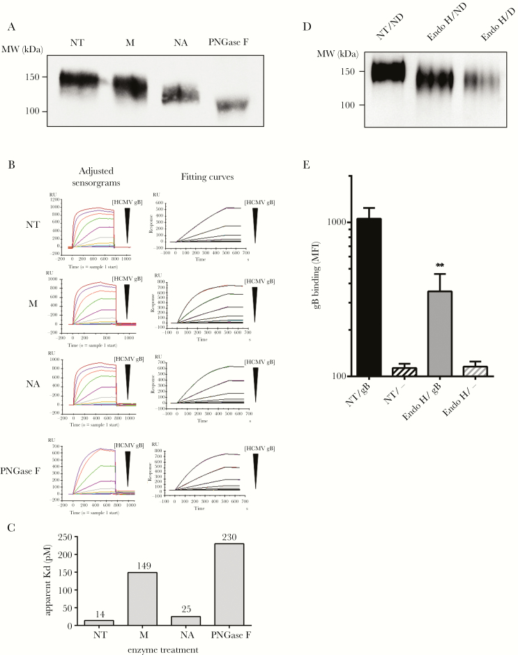
Methods: We previously showed that gB is a ligand for the C-type lectin dendritic cell-specific intercellular adhesion molecule-3-grabbing nonintegrin (DC-SIGN) contributing to HCMV attachment to and infection of DC-SIGN-expressing cells. However, the features of the DC-SIGN/gB interaction remain unclear. To address this point, the role of glycans on gB and the consequences of mutagenesis and antibody-mediated blockades on both partners were examined in this study.

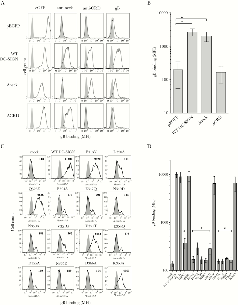
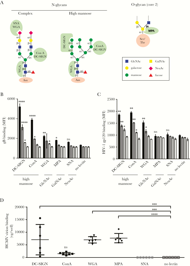
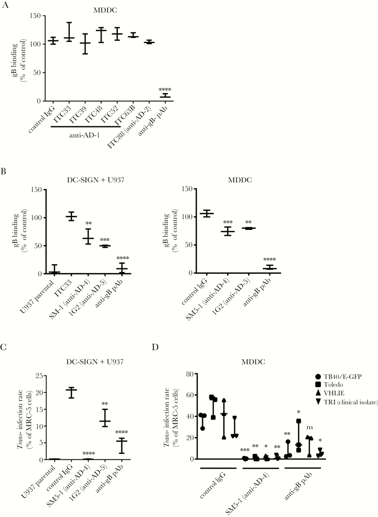
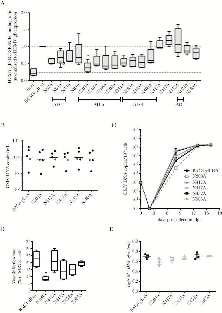
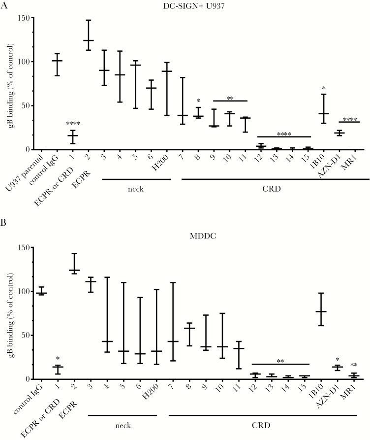
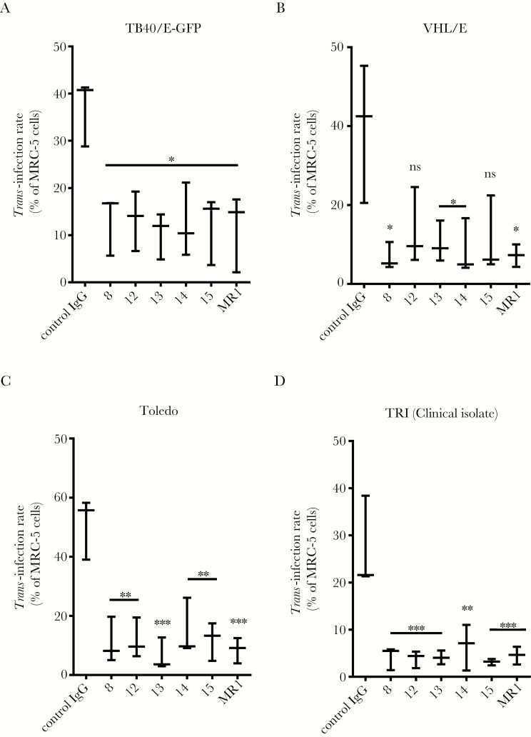
Results: We identified DC-SIGN amino acid residues involved in this interaction through an extensive mutagenesis study. We also showed the importance of high-mannose N-glycans decorating the asparagine residue at position 208, demonstrating that the antigenic domain 5 on gB is involved in the interaction with DC-SIGN. Finally, antibody-mediated blockades allowed us to identify DC-SIGN as a major HCMV attachment receptor on monocyte-derived dendritic cells.

Conclusions: Taken together, these results have permitted us to fine-map the interaction between DC-SIGN and HCMV gB.


Figures

Figure 1.
Figure 1.
Dendritic cell-specific intercellular adhesion molecule-3-grabbing nonintegrin (DC-SIGN) binds the glycoprotein B (gB) through its carbohydrate recognition domain. (A) Histograms showing DC-SIGN expression of wild-type (WT) DC-SIGN or deletion mutants lacking the DC-SIGN neck repeat (Δneck) or the carbohydrate-recognition domain ([CRD] ΔCRD) regions fused to enhanced green fluorescent protein (eGFP). The eGFP allowed a rapid quantitation of the DC-SIGN expression level on stably transfected HEK293T (left panels), except for the pEGFP-transfected cells (first line). The 2 centered columns represent extracellular staining of DC-SIGN with an antineck (clone H-200) and an anti-CRD (clone 1B10) antibody, respectively. The ability of DC-SIGN variants to bind recombinant biotinylated human cytomegalovirus (HCMV) gB is represented in right panels. Gray histograms display nontransfected HEK293T cell fluorescence background. (B) Quantitative measurements of the binding of recombinant biotinylated HCMV gB (2 µg/mL) onto WT DC-SIGN or Δneck- and ΔCRD-expressing cells compared with a control cell line (pEGFP). Biotinylated HCMV gB was revealed with 1 µg/mL antigen-presenting cell-conjugated streptavidin. Values are expressed as mean fluorescence intensities (n = 4; *, P < .05; one-way analysis of variance [ANOVA] with multiple comparison tests). (C) Histograms showing the binding of recombinant Alexa Fluor 647-conjugated HCMV gB (4 µg/mL, mean fluorescence intensity [MFI]) on HEK293T cell lines expressing WT or mutated DC-SIGN on their surface. Values indicated for each histogram represent MFI. These results are representative of 3 independent experiments. (D) Quantitative results showing the behavior of mutated DC-SIGN compared with the WT form towards the binding of recombinant Alexa Fluor 647-conjugated HCMV gB (n = 3). Statistically significant results were marked by an asterisk (*, P < .05; one-way ANOVA with multiple comparison tests).
Figure 2.
Figure 2.
Recombinant soluble or particle-associated human cytomegalovirus (HCMV) glycoprotein B (gB) preferentially interacts with high mannose-specific and N-glycan-specific lectins. (A) Schematic representation of typical N- or O-glycans potentially harbored by glycoproteins. Sugar linkage is indicated between sugar residues. Green zones represent lectin specificities. Lectin binding assay revealing interactions between Alexa 488-conjugated gB (B) or human immunodeficiency virus (HIV)-1 IIIB gp120 (C) and various recombinant plant and animal lectins of known specificities immobilized on plastic 96-well plates. Serial dilutions of each viral glycoprotein were used in this assay: 10 µg/mL (black), 2.5 µg/mL (dark gray), 0.4 µg/mL (light gray), and no gB (open bars). Lectin specificities are indicated beneath each lectin. Results are shown as mean fluorescence intensity ([MFI] n = 3). The “no lectin” condition was used for both recombinant glycoproteins as a negative control providing a basal level of unspecific binding for fluorescence-labeled glycoproteins. (D) Binding of HCMV infectious particles (TB40/E strain, BAC4) to plastic-immobilized plant and animal lectins. A single input of virus was equivalent to 8 × 10⁶ viral genomes/well (vg/well). Results are expressed as vg/well (n = 6). Mean values are represented by horizontal bars for each type of lectin (****, P < .0001; ***, P < .001; **, P < .01; *, P < .05; one-way analysis of variance with multiple comparison tests). Abbreviations: DC-SIGN, dendritic cell-specific intercellular adhesion molecule-3-grabbing nonintegrin; ConA, concanavalin A; MPA, Maclura pomifera agglutinin; ns, nonsignificant; SNA, Sambucus nigra agglutinin; WGA, wheat germ agglutinin.
Figure 3.
Figure 3.
High mannose-containing N-glycans on glycoprotein B (gB) are crucial for its interaction with dendritic cell-specific intercellular adhesion molecule-3-grabbing nonintegrin (DC-SIGN). (A) Western blot analysis showing the detection of recombinant soluble gB with or without enzymatic treatments to remove specifically particular glycan residues. M, NA, and PNGase F stand for the α (1-2,3,6)-mannosidase, the α (2 to 3,6)-neuraminidase, and the peptide-N-glycosidase F, respectively. (B) Adjusted sensorgrams and fitting curves (kinetic model 1:1 binding) for Kd app (apparent Kd) measurements. Black triangles indicate decreasing human cytomegalovirus (HCMV) gB concentration ([HCMV gB]) with 10-fold dilution steps. (C) The affinity of HCMV gB for the extracellular domain of DC-SIGN was determined by surface plasmon resonance (Kd app) before and after deglycosylation with Man, Neu, or PNGase F. (D) Western blot analysis showing from left to right untreated gB, Endo Hf-digested gB in nondenaturing conditions (ND; O/N, 37°C, no denaturation buffer), and Endo Hf-digested gB in denaturing conditions, ie, 1 hour at 37°C with denaturation buffer. (E) Biotinylated HCMV gB was treated with Endo Hf in nondenaturing conditions or left untreated (NT) and further incubated with wild-type DC-SIGN-expressing HEK293T cells. The HCMV gB fixation was revealed as described in the Supplementary Materials. Abbreviation: MW, molecular weight.
Figure 4.
Figure 4.
Antigenic domains (AD)-4- and AD-5- but not AD-1/2-specific antibodies inhibit the dendritic cell-specific intercellular adhesion molecule-3-grabbing nonintegrin (DC-SIGN)/human cytomegalovirus (HCMV) glycoprotein B (gB) interaction and the DC-SIGN-dependent trans-infection of various HCMV strains. (A) Anti-AD-1 (ITC33, ITC39, ITC48, ITC52, and ITC63B) monoclonal antibodies (mAbs), anti-AD-2 (ITC88) mAbs, or a polyclonal anti-HCMV gB serum were used as potential competitors of the HCMV gB binding to immature DC-SIGN+ monocyte-derived dendritic cells (MDDCs). Binding of HCMV gB was assessed by flow cytometry. Binding intensities are represented as mean percentages of the maximum binding (n = 3), ie, without any antibody, and compared with a control immunoglobulin (Ig)G. (B) Inhibition of gB binding on DC-SIGN+ U937 cells compared with the parental counterpart (left panel) or on day 6 immature MDDCs (right panel) by anti-AD-4 (clone SM5-1), anti-AD-5 (clone 1G2), and an anti-gB polyclonal antibody (pAb). Binding intensities are represented as mean percentages of the maximum binding, ie, without any antibody (n = 5). (C) Graph showing trans-infection results obtained with parental vs DC-SIGN+ U937 cells and the TB40/E-GFP strain (multiplicity of infection [MOI] = 2). (D) Similar trans-infection experiments with MDDCs loaded with various low-passage HCMV strains (VHL/E, Toledo, and TB40/E) and a clinical isolate (TRI) (MOI = 2). Asterisks represent significant results compared with the control IgG condition (****, P < .0001; ***, P < .001; **, P < .01; *, P < .05; one-way analysis of variance with multiple comparison tests). Abbreviation: ns, nonsignificant.
Figure 5.
Figure 5.
Mutating the asparagine into an alanine at the position 208 of the human cytomegalovirus (HCMV) gBAA sequence selectively impairs both the HCMV glycoprotein B (gB)/dendritic cell-specific intercellular adhesion molecule-3-grabbing nonintegrin (DC-SIGN) interaction and the DC-SIGN-dependent HCMV trans-infection. (A) Quantitative assessment of the interaction of DC-SIGN-Fc (2 µg/mL) on membrane-immobilized cell lysates of HCMV gB either mutated or wild-type (WT)-expressing HEK293T cells (n = 5). Mean ratios measuring the interaction between WT HCMV gB and DC-SIGN-Fc were normalized to 1 (top gray dashed line), whereas the “mock” condition determines unspecific DC-SIGN-Fc binding on cells transfected with an empty pRC-CMV plasmid (bottom gray dashed line). (B) Five independent viral supernatants were reconstituted for BAC4 gB wt and its mutated versions, ie, N208A, N417A, N447A, N452A, and N585A from BAC-transfected MRC-5 (see Supplementary Materials). The CMV deoxyribonucleic acid (DNA) copy quantification per volume unit by quantitative polymerase chain reaction (qPCR) for all stocks produced in this work is shown here. Grey dots represent stocks from 1 reconstitution. (C) The growth kinetics of the parental HCMV strain (gB WT) or the virus variants with the indicated mutations in the gB protein on MRC5 cells was assessed at day 2, 8, 13, and 16 postinfection. MRC-5 (105) cells were initially infected with a viral dose equivalent to 103 genome copies. Virus amounts are given as genome copies per 105 cells. (D) Wild-type (BAC4 gB wt) and mutated virions (N208A, N417A, N447A, N452A, and N585A) were used to load monocyte-derived dendritic cells (multiplicity of infection [MOI] = 2) for trans-infection experiments. Trans-infection rates represent the percentages of HCMV-infected MRC-5 cells, ie, IE/E antigen-positive among total MRC-5 cells stained at day 2 postinfection (*, P < .05; one-way analysis of variance with multiple comparison tests; n = 4). Results are displayed as a minimum to maximum representation; the box plots show the 10th percentile, the mean (intermediate bars in boxes), and the 90th percentile. (E) In parallel, we collected supernatants of MCR-5 infected with the same amount of virus compared with the one used in trans-infection experiments (MOI = 2). Quantifications by qPCR (CMV deoxyribonucleic acid [DNA] copies/mL) of these supernatants are shown here (n = 3 except for the N585A supernatant where n = 2).
Figure 6.
Figure 6.
Anti-dendritic cell-specific intercellular adhesion molecule-3-grabbing nonintegrin (DC-SIGN) carbohydrate-recognition domain (CRD) antibodies abrogate the DC-SIGN/human cytomegalovirus (HCMV) glycoprotein B (gB) interaction. The binding of conjugated gB (2 µg/mL) to (A) parental or DC-SIGN+ U937 cells or (B) immature monocyte-derived dendritic cells (MDDC) was assessed by flow cytometry. Fifteen available anti-DC-SIGN antibodies directed against the neck, extracellular proximal region (ECPR), or CRD regions were compared with the H200 (anti-neck), 1B10, AZN-D1, and MR-1 (anti-CRD) commercially available monoclonal antibodies for their ability to block the DC-SIGN/HCMV gB interaction. Binding intensities are represented as mean percentages of the maximum binding (n = 5), ie, in the absence of any antibody, and compared with treatment with an irrelevant control immunoglobulin (Ig)G. Asterisks represent significant results (****, P < .0001; **, P < .01; *, P < .05; one-way analysis of variance with multiple comparison tests).
Figure 7.
Figure 7.
Blocking dendritic cell-specific intercellular adhesion molecule-3-grabbing nonintegrin (DC-SIGN) carbohydrate-recognition domain (CRD) strongly impairs human cytomegalovirus (HCMV) trans-infection by monocyte-derived dendritic cells (MDDC). (A) Antibody-mediated blockade of HCMV trans-infection by MDDC. Six anti-DC-SIGN CRD antibodies were used to interfere with the capture and subsequent transmission to highly permissive MRC-5 cells. Monoclonal antibodies were used to block the capture and further transmission of HCMV (TB40/E-GFP strain, multiplicity of infection = 2). Similar experiments showing the trans-infection of the VHL/E (B) and Toledo (C) strains as well as a clinical isolate from our research center, TRI (D). Asterisks represent significant results compared with the control immunoglobulin (Ig)G condition (***, P < .001; **, P < .01; *, P < .05; one-way analysis of variance with multiple comparison tests). Abbreviation: ns, nonsignificant.

Similar articles

Cited by

References

    1. Dioverti MV, Razonable RR. Cytomegalovirus. Microbiol Spectr 2016; 4:Aug; 4(4). - PubMed
    1. Kabanova A, Perez L, Lilleri D et al. Antibody-driven design of a human cytomegalovirus gHgLpUL128L subunit vaccine that selectively elicits potent neutralizing antibodies. Proc Natl Acad Sci U S A 2014; 111:17965–70. - PMC - PubMed
    1. Wussow F, Chiuppesi F, Martinez J et al. Human cytomegalovirus vaccine based on the envelope gH/gL pentamer complex. PLoS Pathog 2014; 10:e1004524. - PMC - PubMed
    1. Kirchmeier M, Fluckiger AC, Soare C et al. Enveloped virus-like particle expression of human cytomegalovirus glycoprotein B antigen induces antibodies with potent and broad neutralizing activity. Clin Vaccine Immunol 2014; 21:174–80. - PMC - PubMed
    1. Wang Z, La Rosa C, Maas R et al. Recombinant modified vaccinia virus Ankara expressing a soluble form of glycoprotein B causes durable immunity and neutralizing antibodies against multiple strains of human cytomegalovirus. J Virol 2004; 78:3965–76. - PMC - PubMed

Publication types

MeSH terms