

Infectious, inflammatory and 'autoimmune' male factor infertility: how do rodent models inform clinical practice?
- PMID: 29648649
- PMCID: PMC6016649
- DOI: 10.1093/humupd/dmy009
Infectious, inflammatory and 'autoimmune' male factor infertility: how do rodent models inform clinical practice?
Abstract
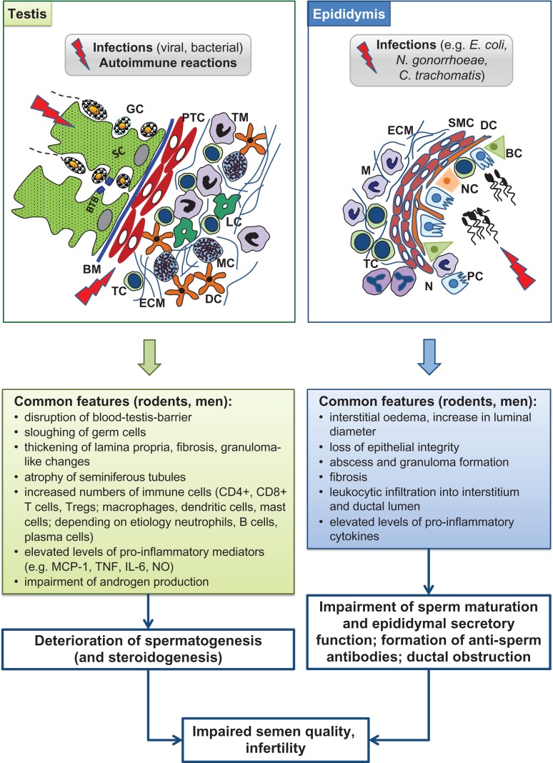
Background: Infection and inflammation of the reproductive tract are significant causes of male factor infertility. Ascending infections caused by sexually transmitted bacteria or urinary tract pathogens represent the most frequent aetiology of epididymo-orchitis, but viral, haematogenous dissemination is also a contributory factor. Limitations in adequate diagnosis and therapy reflect an obvious need for further understanding of human epididymal and testicular immunopathologies and their contribution to infertility. A major obstacle for advancing our knowledge is the limited access to suitable tissue samples. Similarly, the key events in the inflammatory or autoimmune pathologies affecting human male fertility are poorly amenable to close examination. Moreover, the disease processes generally have occurred long before the patient attends the clinic for fertility assessment. In this regard, data obtained from experimental animal models and respective comparative analyses have shown promise to overcome these restrictions in humans.
Objective and rationale: This narrative review will focus on male fertility disturbances caused by infection and inflammation, and the usefulness of the most frequently applied animal models to study these conditions.
Search methods: An extensive search in Medline database was performed without restrictions until January 2018 using the following search terms: 'infection' and/or 'inflammation' and 'testis' and/or 'epididymis', 'infection' and/or 'inflammation' and 'male genital tract', 'male infertility', 'orchitis', 'epididymitis', 'experimental autoimmune' and 'orchitis' or 'epididymitis' or 'epididymo-orchitis', antisperm antibodies', 'vasectomy'. In addition to that, reference lists of primary and review articles were reviewed for additional publications independently by each author. Selected articles were verified by each two separate authors and discrepancies discussed within the team.
Outcomes: There is clear evidence that models mimicking testicular and/or epididymal inflammation and infection have been instructive in a better understanding of the mechanisms of disease initiation and progression. In this regard, rodent models of acute bacterial epididymitis best reflect the clinical situation in terms of mimicking the infection pathway, pathogens selected and the damage, such as fibrotic transformation, observed. Similarly, animal models of acute testicular and epididymal inflammation using lipopolysaccharides show impairment of reproduction, endocrine function and histological tissue architecture, also seen in men. Autoimmune responses can be studied in models of experimental autoimmune orchitis (EAO) and vasectomy. In particular, the early stages of EAO development showing inflammatory responses in the form of peritubular lymphocytic infiltrates, thickening of the lamina propria of affected tubules, production of autoantibodies against testicular antigens or secretion of pro-inflammatory mediators, replicate observations in testicular sperm extraction samples of patients with 'mixed atrophy' of spermatogenesis. Vasectomy, in the form of sperm antibodies and chronic inflammation, can also be studied in animal models, providing valuable insights into the human response.
Wider implications: This is the first comprehensive review of rodent models of both infectious and autoimmune disease of testis/epididymis, and their clinical implications, i.e. their importance in understanding male infertility related to infectious and non-infectious/autoimmune disease of the reproductive organs.
Figures
References
-
- Adams CE, Wald M. Risks and complications of vasectomy. Urol Clin North Am 2009;36:331–336. - PubMed
-
- Adekunle AO, Hickey WF, Smith SM, Tung KS, Teuscher C. Experimental allergic orchitis in mice: IV. Preliminary characterization of the major murine testis specific aspermatogenic autoantigen(s). J Reprod Immunol 1987;12:49–62. - PubMed
-
- Ahmed A, Bello A, Mbibu NH, Maitama HY, Kalayi GD. Epidemiological and aetiological factors of male infertility in northern Nigeria. Niger J Clin Pract 2010;13:205–209. - PubMed
-
- Albrecht M, Frungieri MB, Gonzalez-Calvar S, Meineke V, Kohn FM, Mayerhofer A. Evidence for a histaminergic system in the human testis. Fertil Steril 2005;83:1060–1063. - PubMed
Publication types
MeSH terms
Grants and funding
LinkOut - more resources
Full Text Sources
Other Literature Sources
Medical
