Review
. 2018 May 15;51(5):1046-1053.
doi: 10.1021/acs.accounts.7b00594. Epub 2018 Apr 12.

The Role of Membrane Curvature in Nanoscale Topography-Induced Intracellular Signaling

Affiliations
Review

The Role of Membrane Curvature in Nanoscale Topography-Induced Intracellular Signaling

Hsin-Ya Lou et al. Acc Chem Res. .

Abstract

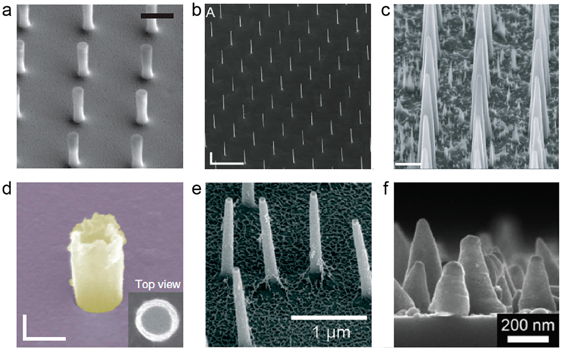
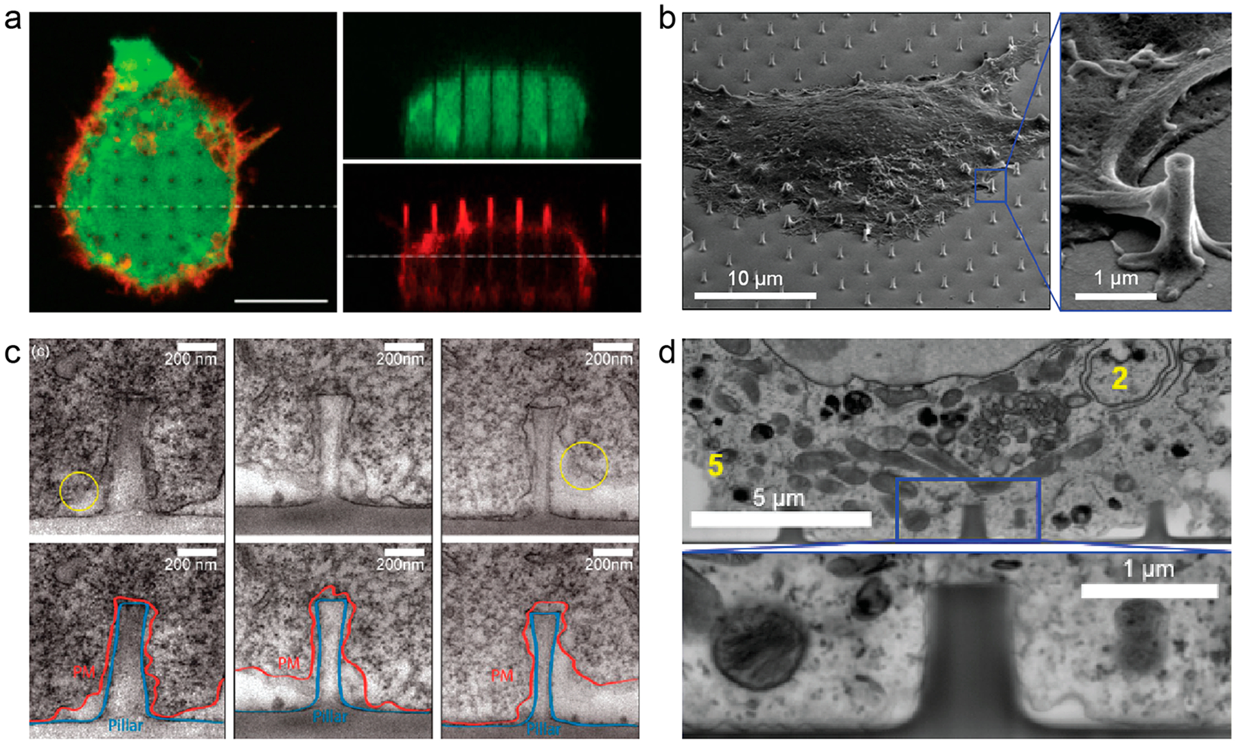
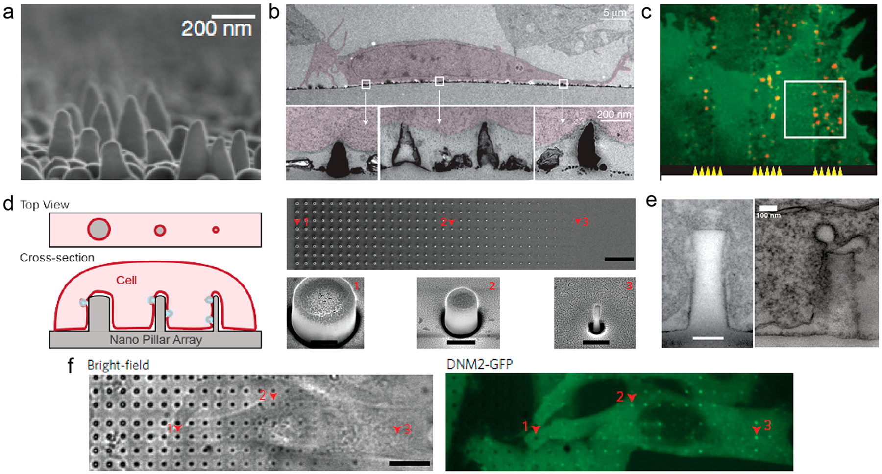
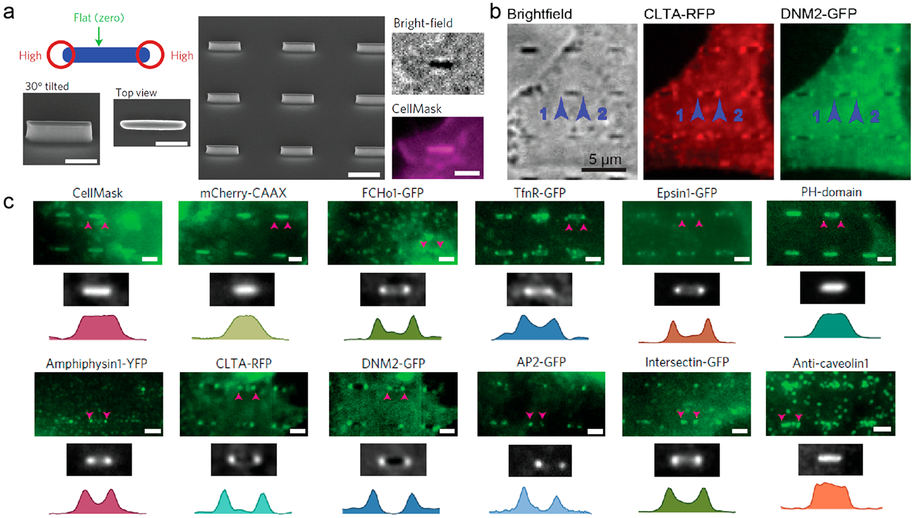
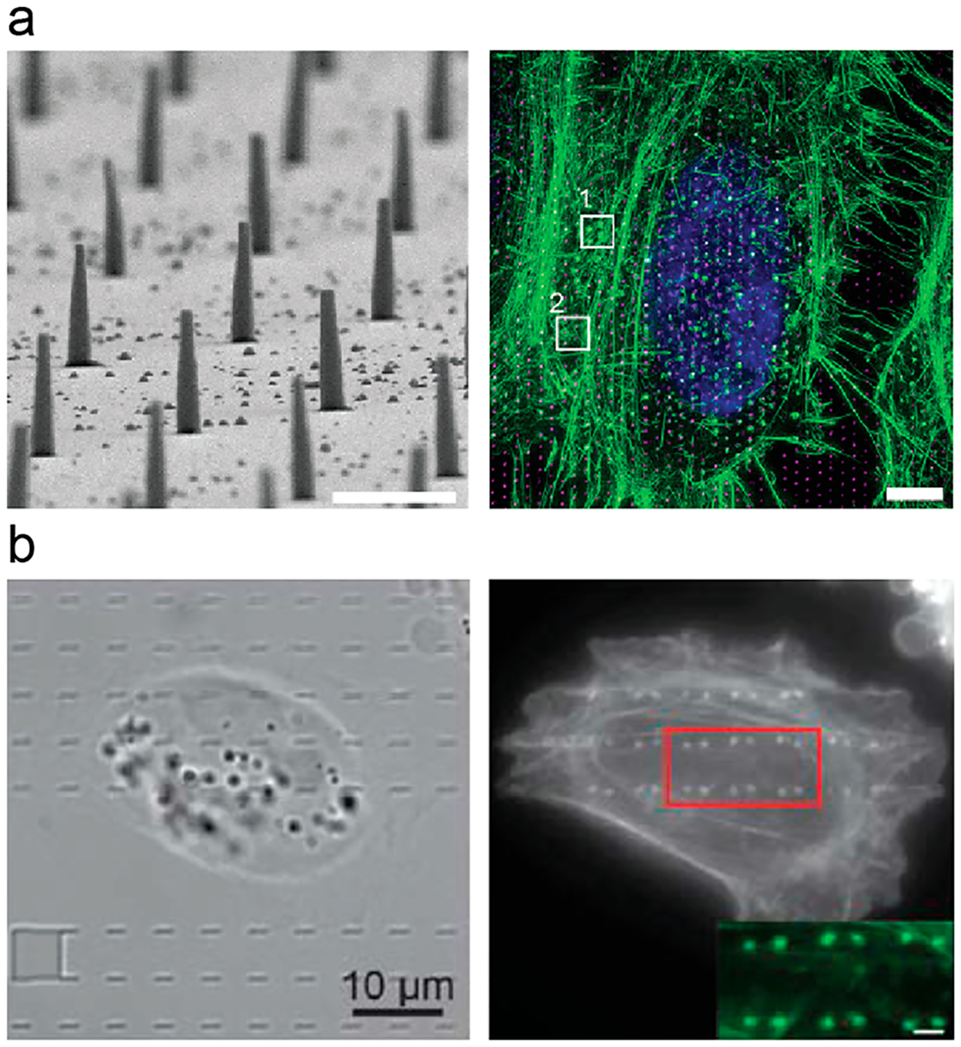
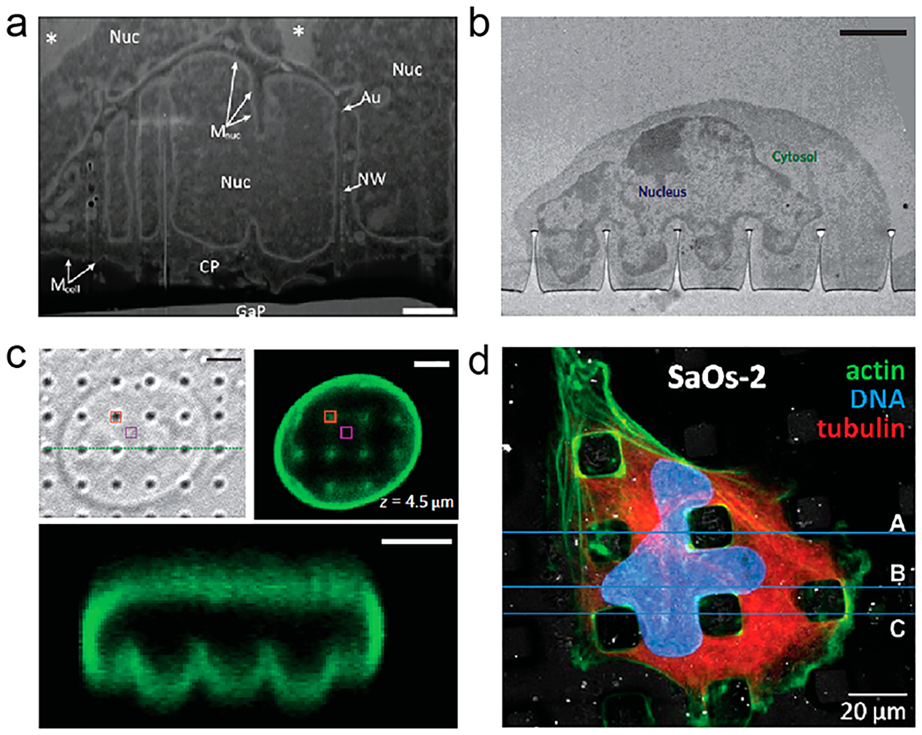
Over the past decade, there has been growing interest in developing biosensors and devices with nanoscale and vertical topography. Vertical nanostructures induce spontaneous cell engulfment, which enhances the cell-probe coupling efficiency and the sensitivity of biosensors. Although local membranes in contact with the nanostructures are found to be fully fluidic for lipid and membrane protein diffusions, cells appear to actively sense and respond to the surface topography presented by vertical nanostructures. For future development of biodevices, it is important to understand how cells interact with these nanostructures and how their presence modulates cellular function and activities. How cells recognize nanoscale surface topography has been an area of active research for two decades before the recent biosensor works. Extensive studies show that surface topographies in the range of tens to hundreds of nanometers can significantly affect cell functions, behaviors, and ultimately the cell fate. For example, titanium implants having rough surfaces are better for osteoblast attachment and host-implant integration than those with smooth surfaces. At the cellular level, nanoscale surface topography has been shown by a large number of studies to modulate cell attachment, activity, and differentiation. However, a mechanistic understanding of how cells interact and respond to nanoscale topographic features is still lacking. In this Account, we focus on some recent studies that support a new mechanism that local membrane curvature induced by nanoscale topography directly acts as a biochemical signal to induce intracellular signaling, which we refer to as the curvature hypothesis. The curvature hypothesis proposes that some intracellular proteins can recognize membrane curvatures of a certain range at the cell-to-material interface. These proteins then recruit and activate downstream components to modulate cell signaling and behavior. We discuss current technologies allowing the visualization of membrane deformation at the cell membrane-to-substrate interface with nanometer precision and demonstrate that vertical nanostructures induce local curvatures on the plasma membrane. These local curvatures enhance the process of clathrin-mediated endocytosis and affect actin dynamics. We also present evidence that vertical nanostructures can induce significant deformation of the nuclear membrane, which can affect chromatin distribution and gene expression. Finally, we provide a brief perspective on the curvature hypothesis and the challenges and opportunities for the design of nanotopography for manipulating cell behavior.


Conflict of interest statement

The authors declare no competing financial interest.

Figures

Figure 1.
Figure 1.
Overview of vertical nanostructures including (a) nanopillars, (b) nanowires, (c) nanoneedles, (d) nanotubes, (e) nanostraws, and (f) nanocones. The diameter, height, and aspect ratio of the vertical nanostructures are as follows: (a) 150 nm, 1.4 μm, 9; (b) 91 nm, 11 μm, 120; (c) 600 nm, 9 μm, 15; (d) 181 nm, 500 nm, 3; (e) 100 nm, 1.5 μm, 15; (f) 50 nm, 200 nm, 4. Scale bars are as follows: (a) 1 μm, (b) 500 nm, (c) 2 μm, (d) 200 nm. Reprinted with permission from the following: ref , Copyright 2015 Nature Publishing Group; ref , Copyright 2012 IOP Publishing Ltd.; ref , Copyright 2015 Nature Publishing Group; ref , Copyright 2014 Nature Publishing Group; ref , Copyright 2016 Royal Society of Chemistry; ref , Copyright 2014 Springer Nature.
Figure 2.
Figure 2.
Vertical nanostructures induce plasma membrane wrapping. Plasma membrane deformation is observed by (a) confocal microscopy (scale bar, 10 μm), (b) SEM, (c) TEM, and (d) FIB/SEM. Reprinted with permission from the following: ref , Copyright 2012 IOP Publishing Ltd.; ref , Copyright 2017 American Chemical Society; ref , Copyright 2012 American Chemical Society.
Figure 3.
Figure 3.
Vertical nanostructures induce local accumulation of proteins. (a) SEM image of the nanocone substrate. (b) TEM images of 3T3 cells on nanocones. (c) Fluorescent images show nadrin-2 (red) preferentially accumulated on nanocone strips compared with flat strips. (d) (left) Schematic illustration of gradient nanopillars deforming the plasma membrane. (right) SEM images of the gradient nanopillars. Scale bars, 10 μm (top), 400 nm (bottom). (e) TEM images of the membrane–nanopillar interface (left) and clathrin-coated pits (right) on the nanopillars. Scale bar, 100 nm. (f) A time-averaged image of dynamin2–GFP demonstrates that dynamin–GFP exhibits strong preference to sharp nanopillars. Scale bar, 10 μm. Reprinted with permission from the following: ref , Copyright 2012 Nature Publishing Group; ref , Copyright 2017 Nature Publishing Group.
Figure 4.
Figure 4.
Clathrin-dependent endocytic proteins accumulate at nanobar ends in a curvature-dependent manner. (a) (left and middle) Schematic and SEM images of nanobar structure. Scale bars, 1 μm. (right) CellMask Deep Red staining of SK-MEL-2 cells on the nanobar arrays. Scale bar, 2 μm. (b) Averaged fluorescence images show that clathrin and dynamin-2 preferentially accumulate around the ends of nanobar structures. Scale bar, 5 μm. (c) The distribution of CellMask, mCherry-CAAX, and various endocytic proteins on the nanobar arrays. Scale bars, 2 μm. Reprinted with permission from ref . Copyright 2017 Nature Publishing Group.
Figure 5.
Figure 5.
Vertical nanostructures induce local polymerization of F-actin. (a) (left) SEM image of SU-8 nanopillar arrays. Scale bar, 1 μm. (right) Maximum intensity projection of a 3D-SIM stack showing phalloidin–Alexa488-labeled F-actin (green), Hoechst 34580 labeled nucleus (blue), and 1 μm spaced nanopillars (magenta) in a hexagonal array. Scale bar, 5 μm. (b) Full frame images of F-actin shows its strong preference on two ends of nanobars, suggesting a curvature effect. Reprinted with permission from the following: ref , Copyright 2015 The Royal Society of Chemistry; ref , Copyright 2017 Nature Publishing Group.
Figure 6.
Figure 6.
Vertical nanostructures induce nuclear membrane deformation. (a, b) TEM images of nuclear membrane deformation on vertical nanowires and nanopillars. Scale bars, (a) 1 μm and (b) 2 μm. (c) Confocal microscopy showed nuclear membrane deformation of 3T3 nucleus on nanopillars. Scale bars, 3 μm. (d) Nuclear deformation between PLLA micropillars showed by fluorescence microscopy. Reprinted with permission from the following: ref , Copyright 2013 Wiley; ref , Copyright 2015 Nature Publishing Group; ref , Copyright 2013 Elsevier.

References

    1. Duan X; Gao R; Xie P; Cohen-Karni T; Qing Q; Choe HS; Tian B; Jiang X; Lieber CM Intracellular Recordings of Action Potentials by an Extracellular Nanoscale Field-Effect Transistor. Nat. Nanotechnol 2012, 7 (3), 174–179. - PMC - PubMed
    1. Xie C; Lin Z; Hanson L; Cui Y; Cui B Intracellular Recording of Action Potentials by Nanopillar Electroporation. Nat. Nanotechnol 2012, 7 (3), 185–190. - PMC - PubMed
    1. Robinson JT; Jorgolli M; Shalek AK; Yoon M-H; Gertner RS; Park H Vertical Nanowire Electrode Arrays as a Scalable Platform for Intracellular Interfacing to Neuronal Circuits. Nat. Nanotechnol 2012, 7 (3), 180–184. - PMC - PubMed
    1. Yang MT; Sniadecki NJ; Chen CS Geometric Considerations of Micro- to Nanoscale Elastomeric Post Arrays to Study Cellular Traction Forces. Adv. Mater 2007, 19 (20), 3119–3123.
    1. Bucaro MA; Vasquez Y; Hatton BD; Aizenberg J Fine-Tuning the Degree of Stem Cell Polarization and Alignment on Ordered Arrays of High-Aspect-Ratio Nanopillars. ACS Nano 2012, 6 (7), 6222–6230. - PubMed

Publication types

MeSH terms

LinkOut - more resources