. 2018 Sep 1;35(17):2067-2076.
doi: 10.1089/neu.2017.5430. Epub 2018 Jun 8.

Traumatic Brain Injury-Induced Acute Lung Injury: Evidence for Activation and Inhibition of a Neural-Respiratory-Inflammasome Axis

Affiliations

Traumatic Brain Injury-Induced Acute Lung Injury: Evidence for Activation and Inhibition of a Neural-Respiratory-Inflammasome Axis

Nadine A Kerr et al. J Neurotrauma. .

Abstract

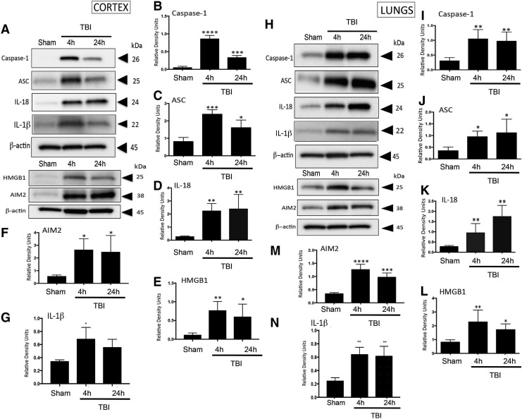
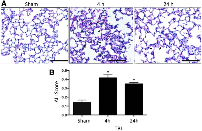
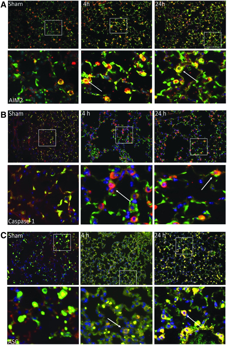
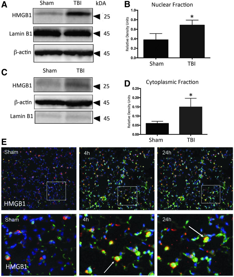
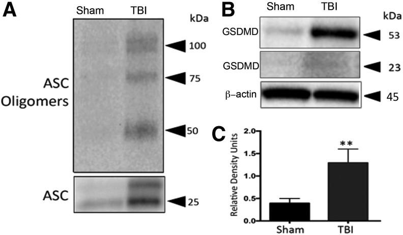
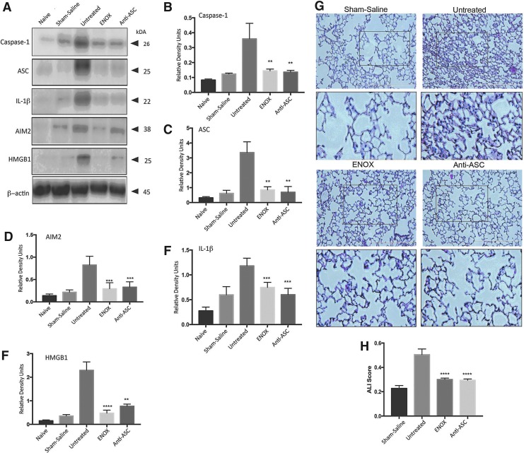
Approximately 20-25% of traumatic brain injury (TBI) subjects develop acute lung injury (ALI), but the pathomechanisms of TBI-induced ALI remain poorly defined. Our previous work has shown that the inflammasome plays a critical role in TBI-induced secondary pathophysiology and that inflammasome proteins are released in extracellular vesicles (EV) after TBI. Here we investigated whether EV-mediated inflammasome signaling contributed to the etiology of TBI-induced ALI. C57/BL6 male mice were subjected to controlled cortical impact (CCI), and the brains and lungs were examined for inflammasome activation and ALI at 4 and 24 h after TBI. We show that TBI releases EV containing inflammasome proteins into serum that target the lung to cause ALI, supporting activation of a neural-respiratory-inflammasome axis. Administration of a low-molecular-weight heparin (enoxaparin, a blocker of EV uptake) or treatment with a monoclonal antibody against apoptosis speck-like staining protein containing a caspase recruitment domain (anti-ASC) after adoptive transfer of EV isolated from TBI-injured mice significantly inhibited inflammasome activation in the lungs of recipient mice resulting in improved ALI scores.This axis constitutes an important arm of the innate inflammatory response in lung pathology after TBI and targeting this axis represents a novel therapeutic treatment for TBI-induced ALI.

Keywords: acute lung injury; extracellular vesicles; inflammasome; innate immune response; traumatic brain injury.


Conflict of interest statement

JPdRV, WDD, and RWK are managing members in InflamaCORE, LLC., a company dedicated to discovering novel diagnostic and therapeutic strategies targeting abnormal inflammasome activation in clinical conditions.

Figures

<b>FIG. 1.</b>
FIG. 1.
Inflammasome activation in C57/BL6 mouse cortical and lung tissue post-TBI. (A) Representative immunoblot of active caspase-1, ASC, IL-18, IL-β, HMGB1, and AIM2 after TBI. (B–G) Active caspase-1, ASC, IL-18, HMGB1, AIM 2, and IL-β, are significantly elevated in cortical tissue at 4 and 24 h post-TBI. Data presented as mean ± SEM; ****p < 0.001, ***p < 0.01, p < 0.05 compared with sham. N = 5–6 per group. (H) Representative immunoblot of active caspase-1, ASC, IL-18, IL-β, HMGB1, and AIM2 in lung tissue. (I–N) Active caspase-1, ASC, IL-18, HMGB1, AIM2, and IL-1β are significantly elevated in lung tissue 4 and 24 h after TBI. Data presented as mean ± SEM. N = 5–6 per group, **p < 0.01., *p < 0.05 compared with sham. ASC, apoptosis speck-like staining protein containing a caspase recruitment domain; IL, interleukin; SEM, standard error of the mean; TBI, traumatic brain injury.
<b>FIG. 2.</b>
FIG. 2.
TBI induces alveolar morphological changes and acute lung injury in mice. (A) H&E staining of lung sections from sham and injured animals at 4 and 24 h. Sections show evidence of neutrophil infiltration (arrow heads), changes in morphology of alveolar capillary membranes (asterisk, *), interstitial edema (short arrows), and evidence of thickening of the interstitium and the alveolar septum (pound, #). (B) Acute lung injury scoring is significantly increased in injured animals when compared with sham at 4 and 24 h. Data presented as mean ± SEM. N = 5–6 per group, **p < 0.01, *p < 0.05 compared with sham. H&E, hematoxylin and eosin; SEM, standard error of the mean; TBI, traumatic brain injury. Color image is available online at www.liebertpub.com/neu
<b>FIG. 3.</b>
FIG. 3.
Expression of inflammasome proteins in type II alveolar epithelial cells. (A) AIM2, (B) active caspase-1, and (C) ASC immunoreactivity increases in lung tissue after CCI (4 h, 24 h) when compared with sham mice. Confocal images of AIM2, caspase-1, and ASC (green signal) and type II epithelial cells (surfactant protein C, red signal), and DAPI nuclear staining (blue signal). Co-localization of inflammasome protein and type II alveolar epithelial cells demonstrated with arrow. ASC, apoptosis speck-like staining protein containing a caspase recruitment domain; CCI, controlled cortical impact. Color image is available online at www.liebertpub.com/neu
<b>FIG. 4.</b>
FIG. 4.
TBI increases nuclear and cytoplasmic HMGB1 expression in mice lung tissue. (A) Representative immunoblot of nuclear HMGB1 after TBI. (B) Nuclear HMGB1 is significantly elevated in 4-h injured animals compared with sham. (C) Representative immunoblot of cytoplasmic HMGB1 after TBI. (D) Cytoplasmic HMGB1 is significantly elevated in 4-h injured animals compared with sham. Data presented as mean ± SEM; ****p < 0.001, ***p < 0.01, *p < 0.05 compared with sham. N = 5–6 per group. (E) HMGB1 immunoreactivity increases in lung tissue after CCI when compared with sham mice. Confocal images of HMGB1(green signal) and type II epithelial cells (surfactant protein C, red signal), and DAPI nuclear staining (blue signal). Co-localization of inflammasome protein and type II alveolar epithelial cells demonstrated with arrow. CCI, controlled cortical impact; SEM, standard error of the mean; TBI, traumatic brain injury. Color image is available online at www.liebertpub.com/neu
<b>FIG. 5.</b>
FIG. 5.
Pyroptosome formation in mice lungs 4 h post-TBI. (A) TBI induces laddering of ASC in lung tissue, indicating formation of the pyroptosome, an oligomerization of ASC dimers that leads to activation of caspase-1 and pyroptosis. (B) Representative immunoblot and (C) quantification of gasdermin. Gasdermin-D is significantly elevated in lung tissue post-TBI. Data presented as mean ± SEM. N = 5 per group, **p < 0.01, *p < 0.05 compared with sham. ASC, apoptosis speck-like staining protein containing a caspase recruitment domain; SEM, standard error of the mean; TBI, traumatic brain injury.
<b>FIG. 6.</b>
FIG. 6.
Treatment with enoxaparin and anti-ASC antibody reduces inflammasome expression in lungs of animals treated with EV from TBI-injured mice. (A) Representative immunoblot showing that caspase-1, ASC, IL-1β, AIM2, HMGB1 are reduced in the lungs of animals that were treated with enoxaparin (3 mg/kg) and IC 100 (5 mg/kg) when compared with untreated positive control animals; (B–F) data presented as mean ± SEM; p < .0.05 compared with sham. N = 4–5 per group. (G) H&E staining of lung sections from saline, untreated, enoxaparin and IC 100 treated mice lungs delivered EV from injured animals. Sections show evidence of neutrophil infiltration, changes in morphology of alveolar capillary membranes, interstitial edema, and evidence of thickening of the interstitium and the alveolar septum. (H) Acute lung injury scoring is significantly decreased in animals treated with enoxaparin or anti-ASC antibody when compared with untreated animals. Data presented as mean ± SEM. N = 4–5 per group, **p < 0.01, *p < 0.05. ASC, apoptosis speck-like staining protein containing a caspase recruitment domain; EV, extracellular vesicles; H&E, hematoxylin and eosin; IL, interleukin; SEM, standard error of the mean; TBI, traumatic brain injury. Color image is available online at www.liebertpub.com/neu

Similar articles

Cited by

References

    1. Summers C.R., Ivins B., and Schwab K.A. (2009). Traumatic brain injury in the United States: an epidemiologic overview. Mt. Sinai J. Med. 76, 105–110 - PubMed
    1. Bramlett H.M., and Dietrich W.D. (2015). Long-term consequences of traumatic brain injury: current status of potential mechanisms of injury and neurological outcomes. J. Neurotrauma 32, 1834–1848 - PMC - PubMed
    1. Chovatiya R., and Medzhitov R. (2014). Stress, inflammation, and defense of homeostasis. Mol. Cell 54, 281–288 - PMC - PubMed
    1. Newton K., and Dixit V.M. (2012). Signaling in innate immunity and inflammation. Cold Spring Harb. Perspect. Bio. 4 pii: a006049 - PMC - PubMed
    1. de Rivero Vaccari J.P., Dietrich W.D., and Keane R.W. (2014). Activation and regulation of cellular inflammasomes: gaps in our knowledge for central nervous system injury. J. Cereb. Blood Flow Metab. 34, 369–375 - PMC - PubMed

Publication types

MeSH terms

LinkOut - more resources