

A Single Primary Blast-Induced Traumatic Brain Injury in a Rodent Model Causes Cell-Type Dependent Increase in Nicotinamide Adenine Dinucleotide Phosphate Oxidase Isoforms in Vulnerable Brain Regions
- PMID: 29648986
- PMCID: PMC6098412
- DOI: 10.1089/neu.2017.5358
A Single Primary Blast-Induced Traumatic Brain Injury in a Rodent Model Causes Cell-Type Dependent Increase in Nicotinamide Adenine Dinucleotide Phosphate Oxidase Isoforms in Vulnerable Brain Regions
Abstract
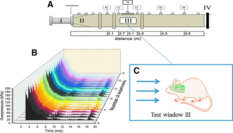
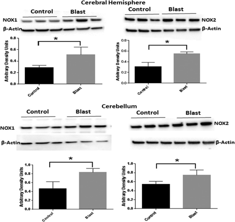
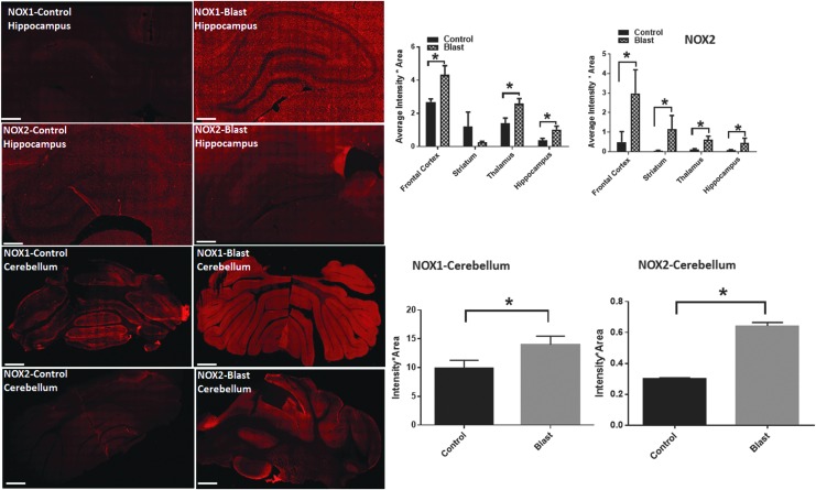
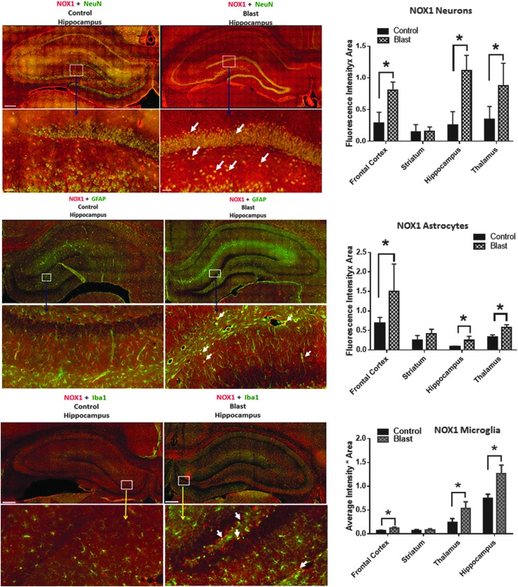
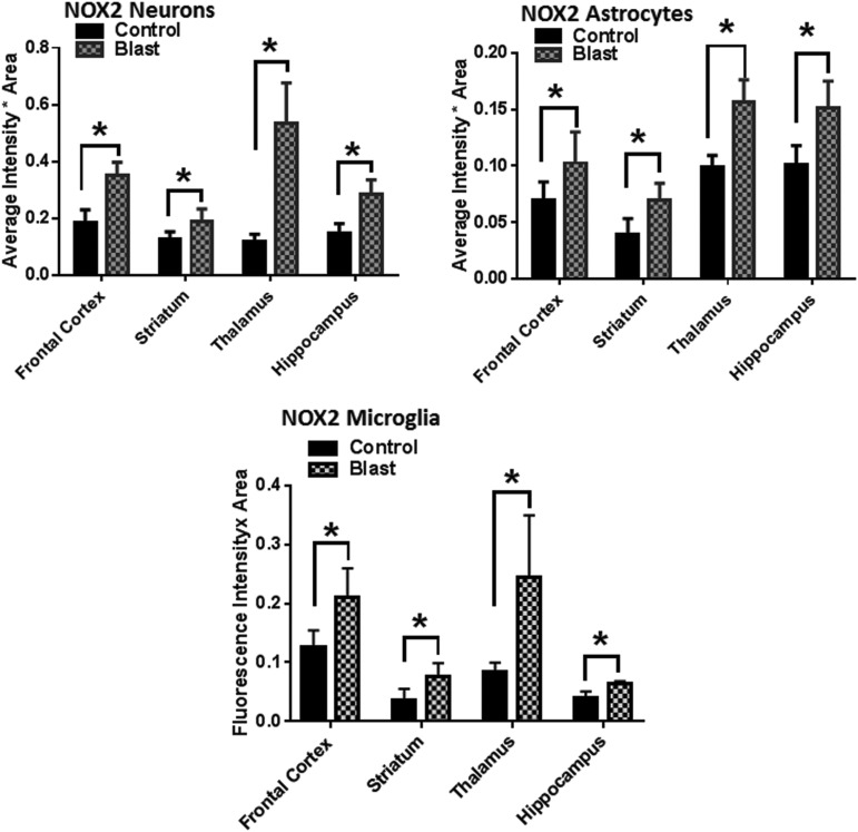
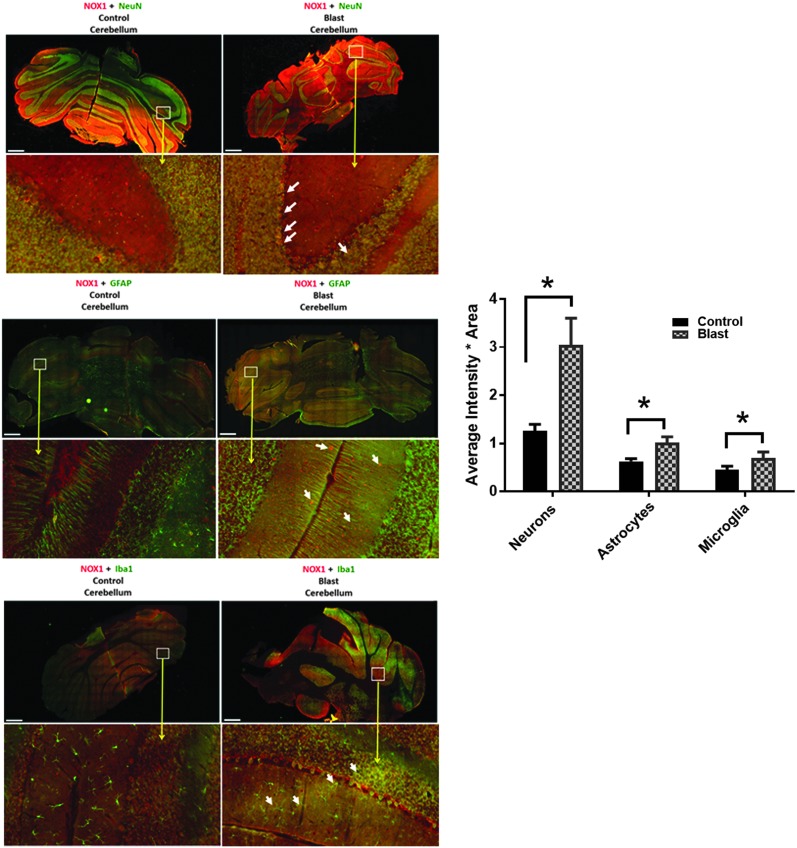
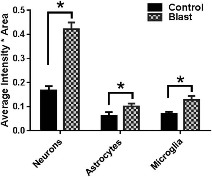
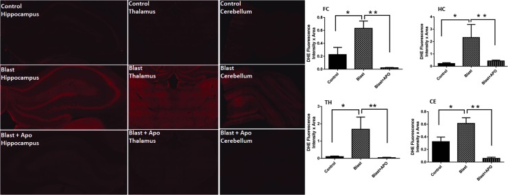
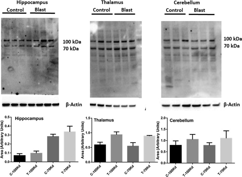
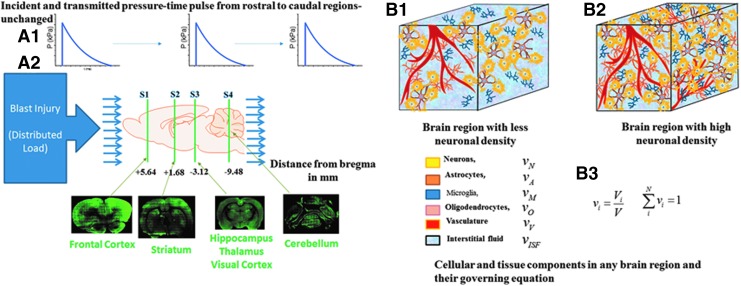
Blast-induced traumatic brain injury (bTBI) is a leading cause of morbidity in soldiers on the battlefield and in training sites with long-term neurological and psychological pathologies. Previous studies from our laboratory demonstrated activation of oxidative stress pathways after blast injury, but their distribution among different brain regions and their impact on the pathogenesis of bTBI have not been explored. The present study examined the protein expression of two isoforms: nicotinamide adenine dinucleotide phosphate (NADPH) oxidase 1 and 2 (NOX1, NOX2), corresponding superoxide production, a downstream event of NOX activation, and the extent of lipid peroxidation adducts of 4-hydroxynonenal (4HNE) to a range of proteins. Brain injury was evaluated 4 h after the shock-wave exposure, and immunofluorescence signal quantification was performed in different brain regions. Expression of NOX isoforms displayed a differential increase in various brain regions: in hippocampus and thalamus, there was the highest increase of NOX1, whereas in the frontal cortex, there was the highest increase of NOX2 expression. Cell-specific analysis of changes in NOX expression with respect to corresponding controls revealed that blast resulted in a higher increase of NOX1 and NOX 2 levels in neurons compared with astrocytes and microglia. Blast exposure also resulted in increased superoxide levels in different brain regions, and such changes were reflected in 4HNE protein adduct formation. Collectively, this study demonstrates that primary blast TBI induces upregulation of NADPH oxidase isoforms in different regions of the brain parenchyma and that neurons appear to be at higher risk for oxidative damage compared with other neural cells.
Keywords: 4-hydroxynonenal; NADPH oxidase; astrocytes; blast injury; microglia; neuron; oxidative stress; traumatic brain injury.
Conflict of interest statement
No competing financial interests exist.
Figures
Similar articles
-
Oxidative stress contributes to cerebral metabolomic profile changes in animal model of blast-induced traumatic brain injury.Metabolomics. 2020 Mar 12;16(3):39. doi: 10.1007/s11306-020-1649-4. Metabolomics. 2020. PMID: 32166461
-
Synergistic Role of Oxidative Stress and Blood-Brain Barrier Permeability as Injury Mechanisms in the Acute Pathophysiology of Blast-induced Neurotrauma.Sci Rep. 2019 May 22;9(1):7717. doi: 10.1038/s41598-019-44147-w. Sci Rep. 2019. PMID: 31118451 Free PMC article.
-
Amelioration of nicotinamide adenine dinucleotide phosphate-oxidase mediated stress reduces cell death after blast-induced traumatic brain injury.Transl Res. 2015 Dec;166(6):509-528.e1. doi: 10.1016/j.trsl.2015.08.005. Epub 2015 Sep 8. Transl Res. 2015. PMID: 26414010
-
NADPH oxidases in traumatic brain injury - Promising therapeutic targets?Redox Biol. 2018 Jun;16:285-293. doi: 10.1016/j.redox.2018.03.005. Epub 2018 Mar 15. Redox Biol. 2018. PMID: 29571125 Free PMC article. Review.
-
Central Nervous System Injury and Nicotinamide Adenine Dinucleotide Phosphate Oxidase: Oxidative Stress and Therapeutic Targets.J Neurotrauma. 2017 Feb 15;34(4):755-764. doi: 10.1089/neu.2016.4486. Epub 2016 Jun 27. J Neurotrauma. 2017. PMID: 27267366 Free PMC article. Review.
Cited by
-
Phylogenetic conservation of the interdependent homeostatic relationship of sleep regulation and redox metabolism.J Comp Physiol B. 2024 Jun;194(3):241-252. doi: 10.1007/s00360-023-01530-4. Epub 2024 Feb 7. J Comp Physiol B. 2024. PMID: 38324048 Free PMC article. Review.
-
Temporal Changes in Functional and Structural Neuronal Activities in Auditory System in Non-Severe Blast-Induced Tinnitus.Medicina (Kaunas). 2023 Sep 18;59(9):1683. doi: 10.3390/medicina59091683. Medicina (Kaunas). 2023. PMID: 37763802 Free PMC article.
-
Oxidative stress contributes to cerebral metabolomic profile changes in animal model of blast-induced traumatic brain injury.Metabolomics. 2020 Mar 12;16(3):39. doi: 10.1007/s11306-020-1649-4. Metabolomics. 2020. PMID: 32166461
-
Matrix Inversion and Subset Selection (MISS): A pipeline for mapping of diverse cell types across the murine brain.Proc Natl Acad Sci U S A. 2022 Apr 5;119(14):e2111786119. doi: 10.1073/pnas.2111786119. Epub 2022 Apr 1. Proc Natl Acad Sci U S A. 2022. PMID: 35363567 Free PMC article.
-
Twenty Years of Blast-Induced Neurotrauma: Current State of Knowledge.Neurotrauma Rep. 2024 Mar 14;5(1):243-253. doi: 10.1089/neur.2024.0001. eCollection 2024. Neurotrauma Rep. 2024. PMID: 38515548 Free PMC article.
References
-
- Bochicchio G.V., Lumpkins K., O'Connor J., Simard M., Schaub S., Conway A., Bochicchio K., and Scalea T.M. (2008). Blast injury in a civilian trauma setting is associated with a delay in diagnosis of traumatic brain injury. Am. Surg. 74, 267–270 - PubMed
-
- DuBose J.J., Barmparas G., Inaba K., Stein D.M., Scalea T., Cancio L.C., Cole J., Eastridge B., and Blackbourne L. (2011). Isolated severe traumatic brain injuries sustained during combat operations: demographics, mortality outcomes, and lessons to be learned from contrasts to civilian counterparts. J. Trauma 70, 11–16 - PubMed
-
- Reid M.W. and Velez C.S. (2015). Discriminating military and civilian traumatic brain injuries. Mol. Cell. Neurosci. 66, 123–128 - PubMed
-
- Wallace D. (2009). Improvised explosive devices and traumatic brain injury: the military experience in Iraq and Afghanistan. Australas. Psychiatry 17, 218–224 - PubMed
Publication types
MeSH terms
Substances
LinkOut - more resources
Full Text Sources
Other Literature Sources
Medical
Research Materials
Miscellaneous