

NUT Carcinoma of the Salivary Glands: Clinicopathologic and Molecular Analysis of 3 Cases and a Survey of NUT Expression in Salivary Gland Carcinomas
- PMID: 29649019
- PMCID: PMC6010176
- DOI: 10.1097/PAS.0000000000001046
NUT Carcinoma of the Salivary Glands: Clinicopathologic and Molecular Analysis of 3 Cases and a Survey of NUT Expression in Salivary Gland Carcinomas
Abstract
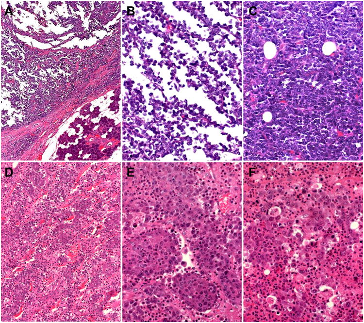
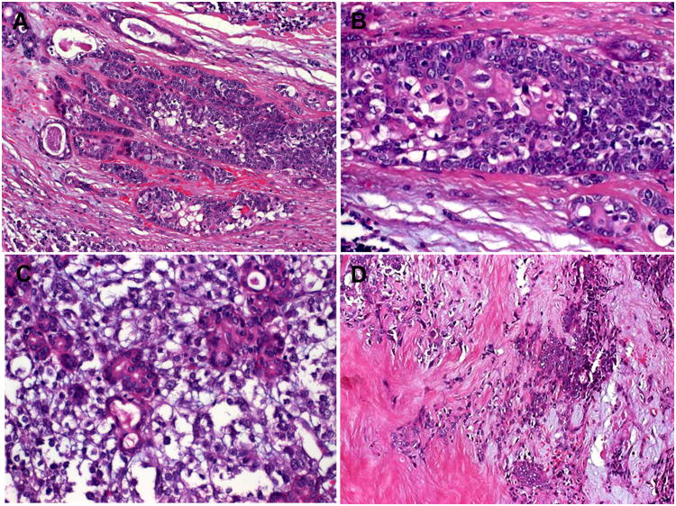
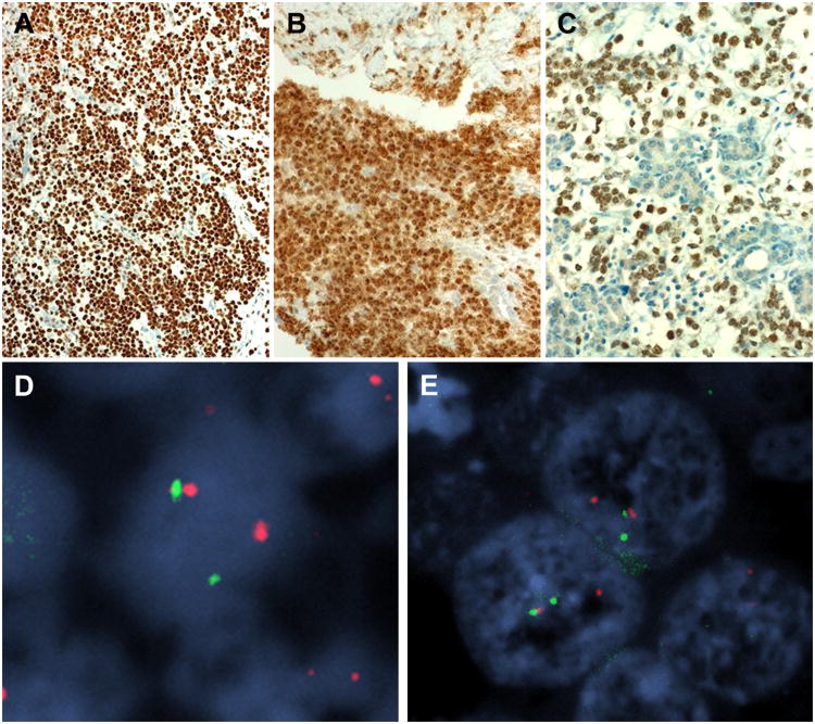
NUT carcinoma (NC) represents a rare subset of highly aggressive poorly differentiated carcinomas characterized by rearrangement of the NUT (aka NUTM1, nuclear protein in testis) gene, most commonly fused to BRD4. Originally described as a mediastinal/thymic malignancy, NC has been reported at a variety of anatomic regions including the upper and lower aerodigestive tract. To date, only 7 NC cases of probable salivary gland origin have been reported. We herein describe 3 new cases (all affecting the parotid gland) in 2 women (39- and 55-y old) and 1 man (35-y old). Histologic examination showed poorly differentiated neoplasms composed of poorly cohesive small-sized to medium-sized cells with variable squamoid cell component that was focal and abrupt. Immunohistochemistry showed uniform expression of p63 and distinctive punctate expression of the NUT antigen in the tumor cell nuclei. Review of the reported salivary gland NC cases (total, 10) showed a male:female ratio of 1.5:1 and an age range of 12 to 55 years (median, 29 y). Site of the primary tumor was the parotid (7), sublingual (2), and submandibular (1) glands. All presented as rapidly growing masses treated by surgery followed by adjuvant radiotherapy/chemotherapy. Initial nodal status was positive in 8/10. At last follow-up (1 to 24 mo; median, 5 mo), 7/10 patients died of disease at a median of 5.5 months (1 to 24 mo) and only 2 were disease free at 7 and 14 months. Of 9 cases with genetic data, the fusion partner was BRD4 (n=7), non-BRD4/3 (n=1), or undetermined (n=1). None of 306 carcinomas spanning the spectrum of salivary carcinoma types screened by NUT immunohistochemistry was positive. This is the first small series on salivary NC highlighting the importance to include this rare disease in the differential diagnosis of poorly differentiated salivary gland carcinomas and in cases of presumable poorly differentiated carcinoma of unknown origin.
Conflict of interest statement
Conflict of interest: none
Figures
References
-
- French CA, Miyoshi I, Kubonishi I, Grier HE, Perez-Atayde AR, Fletcher JA. BRD4-NUT fusion oncogene: a novel mechanism in aggressive carcinoma. Cancer Res. 2003;63:304–7. - PubMed
-
- Kubonishi I, Takehara N, Iwata J, et al. Novel t(15;19)(q15;p13) chromosome abnormality in a thymic carcinoma. Cancer Res. 1991;51:3327–3328. - PubMed
-
- French C. NUT midline carcinoma. Nat Rev Cancer. 2014;14:149–150. - PubMed
-
- French CA, Kutok JL, Faquin WC, et al. Midline carcinoma of children and young adults with NUT rearrangement. J Clin Oncol. 2004;22:4135–4139. - PubMed
-
- Stelow EB, French CA. Carcinomas of the upper aerodigestive tract with rearrangement of the nuclear protein of the testis (NUT) gene (NUT midline carcinomas) Adv Anat Pathol. 2009;16:92–6. - PubMed
Publication types
MeSH terms
Substances
Grants and funding
LinkOut - more resources
Full Text Sources
Other Literature Sources
