. 2018 Apr 12;10(4):473.
doi: 10.3390/nu10040473.

Coordination of GPR40 and Ketogenesis Signaling by Medium Chain Fatty Acids Regulates Beta Cell Function

Affiliations

Coordination of GPR40 and Ketogenesis Signaling by Medium Chain Fatty Acids Regulates Beta Cell Function

Julien Benjamin Pujol et al. Nutrients. .

Abstract

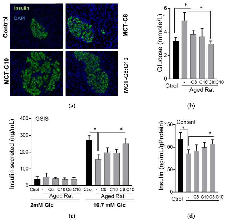
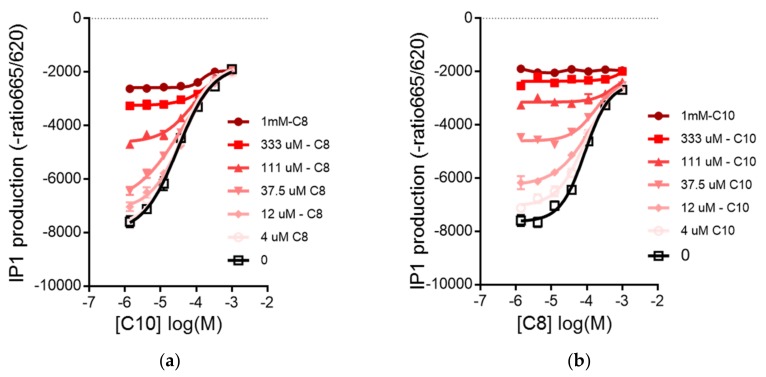
Diabetes prevalence increases with age, and β-cell dysfunction contributes to the incidence of the disease. Dietary lipids have been recognized as contributory factors in the development and progression of the disease. Unlike long chain triglycerides, medium chain triglycerides (MCT) increase fat burning in animal and human subjects as well as serum C-peptide in type 2 diabetes patients. We evaluated the beneficial effects of MCT on β-cells in vivo and in vitro. MCT improved glycemia in aged rats via β-cell function assessed by measuring insulin secretion and content. In β-cells, medium chain fatty acid (MCFA)-C10 activated fatty acid receptor 1 FFAR1/GPR40, while MCFA-C8 induced mitochondrial ketogenesis and the C8:C10 mixture improved β cell function. We showed that GPR40 signaling positively impacts ketone body production in β-cells, and chronic treatment with β-hydroxybutyrate (BHB) improves β-cell function. We also showed that BHB and MCFA help β-cells recover from lipotoxic stress by improving mitochondrial function and increasing the expression of genes involved in β-cell function and insulin biogenesis, such as Glut2, MafA, and NeuroD1 in primary human islets. MCFA offers a therapeutic advantage in the preservation of β-cell function as part of a preventative strategy against diabetes in at risk populations.

Keywords: BHB; GPR40; insulin secretion; ketogenesis; lipotoxicity; medium chain fatty acid; mitochondria.


Conflict of interest statement

The authors declare no conflict of interest.

Figures

Figure 1
Figure 1
Medium chain fatty acids improved β-cell function in aged rats. (a) Immunohistochemical staining of pancreas with insulin from aged rats fed with normal chow diet supplemented with sunflower oil (Ctrol), octanoic acid (C8), decanoic acid (C10) or a 40:60 mixture of octanoic acid: decanoic acid (C8:C10) medium chain triglycerides for 8 weeks. (b) Fasting glucose (n = 12), (c) i secretion and (d) insulin content of young rats (3 months old) compared to aged rats (18 months old) treated as above after 16 h fasting (n = 6, * p < 0.05).
Figure 2
Figure 2
Activation of GPR40 by medium chain fatty acids, C8 and C10, in Chem1-GPR40 cells. (a) Dose–response curves of C8 in the presence of C10; dose–response of IP1 production with C8 activation in the presence of increasing concentrations of C10 and (b) dose–response curves of C10 in the presence of C8; C10 in the presence of increasing concentrations of C8. (c) Dose–response curves of C8 in GW1100; inhibitory effect of the GPR40 antagonist, GW1100, on the dose–response accumulation of IP1 by C8 or by (d) dose–response curves of C10 in GW1100; C10 in Chem1-GPR40 cells. Curves represent as means + SD of eight experimental replicates.
Figure 2
Figure 2
Activation of GPR40 by medium chain fatty acids, C8 and C10, in Chem1-GPR40 cells. (a) Dose–response curves of C8 in the presence of C10; dose–response of IP1 production with C8 activation in the presence of increasing concentrations of C10 and (b) dose–response curves of C10 in the presence of C8; C10 in the presence of increasing concentrations of C8. (c) Dose–response curves of C8 in GW1100; inhibitory effect of the GPR40 antagonist, GW1100, on the dose–response accumulation of IP1 by C8 or by (d) dose–response curves of C10 in GW1100; C10 in Chem1-GPR40 cells. Curves represent as means + SD of eight experimental replicates.
Figure 3
Figure 3
GPR40 signaling affects fatty acid β-oxidation in β-cells. (a) Ketone body β- hydroxybutyrate (BHB) production in INS1E β-cells in the presence of C8, C10 or C8:C10 40:60 mixture. (b) Inhibition of GPR40 signaling by the antagonist, GW1100 (GW), (c) the phospholipase C-β (PLCβ) inhibitor, U73122 (U7) and nifedipine (Nif) prevent BHB production from medium chain fatty acids (MCFA) in INS1E β-cells unlike the IP3 receptor inhibitor xantospongin (Xant). (d) Inhibition of GPR40 signaling by GW increased complete β-oxidation of palmitate. Data are presented as means + SEM of three independent experiments, * p < 0.05, ** p < 0.01 relative to control.
Figure 3
Figure 3
GPR40 signaling affects fatty acid β-oxidation in β-cells. (a) Ketone body β- hydroxybutyrate (BHB) production in INS1E β-cells in the presence of C8, C10 or C8:C10 40:60 mixture. (b) Inhibition of GPR40 signaling by the antagonist, GW1100 (GW), (c) the phospholipase C-β (PLCβ) inhibitor, U73122 (U7) and nifedipine (Nif) prevent BHB production from medium chain fatty acids (MCFA) in INS1E β-cells unlike the IP3 receptor inhibitor xantospongin (Xant). (d) Inhibition of GPR40 signaling by GW increased complete β-oxidation of palmitate. Data are presented as means + SEM of three independent experiments, * p < 0.05, ** p < 0.01 relative to control.
Figure 4
Figure 4
Regulation of insulin secretion by MCFA and BHB in INS1E cells. (a) Increase of calcium mobilization by C8 and C10. (b) Acute statistic insulin secretion after 1 h of treatment with secretagogues, like 1× amino-acids (A), 0.1 µM Exendin-4 (Ex) and 200 µM fatty acids—palmitate (PA, C16:0), C8, C10, in low (2 mM) and high (16.7 mM) glucose or (c) 200 µM C8:C10 40:60 mixture or 200 µM of BHB. (d) Static insulin secretion after chronic (72 h) treatment with 200 µM of C8:C10 40:60 mixture, BHB or palmitate (P). Data represent means + SEM of three independent experiments, * p < 0.05, ** p < 0.01 relative to control.
Figure 4
Figure 4
Regulation of insulin secretion by MCFA and BHB in INS1E cells. (a) Increase of calcium mobilization by C8 and C10. (b) Acute statistic insulin secretion after 1 h of treatment with secretagogues, like 1× amino-acids (A), 0.1 µM Exendin-4 (Ex) and 200 µM fatty acids—palmitate (PA, C16:0), C8, C10, in low (2 mM) and high (16.7 mM) glucose or (c) 200 µM C8:C10 40:60 mixture or 200 µM of BHB. (d) Static insulin secretion after chronic (72 h) treatment with 200 µM of C8:C10 40:60 mixture, BHB or palmitate (P). Data represent means + SEM of three independent experiments, * p < 0.05, ** p < 0.01 relative to control.
Figure 5
Figure 5
MCFA helps the β-cells recover from chronic palmitate induced dysfunction. (a) Representative immunoblotting of INS1E cells after chronic treatment with increased concentrations of palmitate or MCFA C8:C10 mixture with antibodies against Nrf2 and mitochondrial respiratory chain proteins. (b) Assessment of apoptotic and ER stress signaling after chronic treatment with palmitate (C16:0) or MCFA C8:C10 mixture after immunoblotting with antibodies against PARP (Poly(ADP-Ribose) Polymerase), CHOP, Cox4 (Cytochrome C Oxidase Subunit 4) and GAPDH (Glyceraldehyde-3-Phosphate Dehydrogenase) as loading controls. (c) Mitochondrial membrane potential (MMP) in response to 16.7 mM glucose in INS-1E cells chronically treated with vehicle control (Ctrl) or the MCFA C8:C10 mixture, using the fluorescent lipophilic cation IP10. Changes in MMP are expressed as the ratio of the fluorescence in mitochondria divided by the cytosolic fluorescence (Fmito/Fcyto) measured in the same cells. At the end of the recording, the protonophore, FCCP (2 μM) was used to dissipate MMP. Data shown are the means ± SEM from three independent experiments done in sextuplet in 96-welled plates, ** p < 0.01. (d) Expression of CHOP and Glut2 by qPCR in INS1E cells after chronic treatment with palmitate and recovery in control medium or MCFA-enriched medium. (e) Western blot analysis of transcription factors regulating β-cell function (MafA, NeuroD1, PDX1) or mitochondrial proteins (NDUFA9, DLD, Cox4) after treatment as above; GAPDH was used as a loading control. (f) Analysis of β-cell function after impairment with chronic palmitate and recovery as above by insulin secretion under basal (2 mM glucose) and stimulated (16.7 mM glucose + 0.1 µM Ex-4) conditions. Data represent means + SEM of three independent experiments; * p < 0.05, ** p < 0.01 relative to control cells and # p < 0.05 relative to palmitate control cells.
Figure 6
Figure 6
Chronic MCFA treatment improves β-cell function in human islets. (a) Glucose stimulated insulin secretion (GSIS) and (b) insulin content in healthy human islets treated with palmitate (P, 0.5 mM for 72 h) followed by recovery in the presence or absence of the medium chain fatty acid mixture, C8:C10 40:60. (c) Fold change of selected genes in human islets treated as above, using a NanoString n-counter gene expression analysis.
Figure 7
Figure 7
Model of MCFA signaling in β-cells. Pancreatic β-cell function was assessed by the capacity of the β-cell to sense and respond to glucose stimulation through uptake by Glut2 and mitochondrial metabolism leading to an increased ATP/ADP. Inhibition of the kATP channel by ATP leads to membrane depolarization and influx of calcium through the l-type calcium channel that triggers insulin secretion. Preferentially, the activation of GPR40 by C10 MCFA leads to PLCβ activation and generation of IP3 induced ER-calcium release and DAG activation of PKC, which amplifies glucose stimulated insulin release. C8, on the other hand, preferentially increases BHB production. GPR40 activation promotes mitochondrial ketogenesis through Ca2+ and certainly through DAG-PKC signaling. The illustration was made by using tools from Servier Medical Art, http://www.servier.fr/ servier-medical-art.

References

    1. Cowie C.C., Rust K.F., Byrd-Holt D.D., Eberhardt M.S., Flegal K.M., Engelgau M.M., Saydah S.H., Williams D.E., Geiss L.S., Gregg E.W. Prevalence of diabetes and impaired fasting glucose in adults in the U.S. population: National Health And Nutrition Examination Survey 1999–2002. Diabetes Care. 2006;29:1263–1268. doi: 10.2337/dc06-0062. - DOI - PubMed
    1. Cheng Y.J., Gregg E.W., Geiss L.S., Imperatore G., Williams D.E., Zhang X., Albright A.L., Cowie C.C., Klein R., Saaddine J.B. Association of A1C and fasting plasma glucose levels with diabetic retinopathy prevalence in the U.S. population: Implications for diabetes diagnostic thresholds. Diabetes Care. 2009;32:2027–2032. doi: 10.2337/dc09-0440. - DOI - PMC - PubMed
    1. Scheen A.J. Diabetes mellitus in the elderly: Insulin resistance and/or impaired insulin secretion? Diabetes Metab. 2005;31:5S27–5S34. doi: 10.1016/S1262-3636(05)73649-1. - DOI - PubMed
    1. Gunasekaran U., Gannon M. Type 2 diabetes and the aging pancreatic beta cell. Aging (Albany NY) 2011;3:565–575. doi: 10.18632/aging.100350. - DOI - PMC - PubMed
    1. Weyer C., Bogardus C., Mott D.M., Pratley R.E. The natural history of insulin secretory dysfunction and insulin resistance in the pathogenesis of type 2 diabetes mellitus. J. Clin. Investig. 1999;104:787–794. doi: 10.1172/JCI7231. - DOI - PMC - PubMed

MeSH terms