

Tumor-Associated Macrophages and Mast Cells Positive to Tryptase Are Correlated with Angiogenesis in Surgically-Treated Gastric Cancer Patients
- PMID: 29649166
- PMCID: PMC5979483
- DOI: 10.3390/ijms19041176
Tumor-Associated Macrophages and Mast Cells Positive to Tryptase Are Correlated with Angiogenesis in Surgically-Treated Gastric Cancer Patients
Abstract
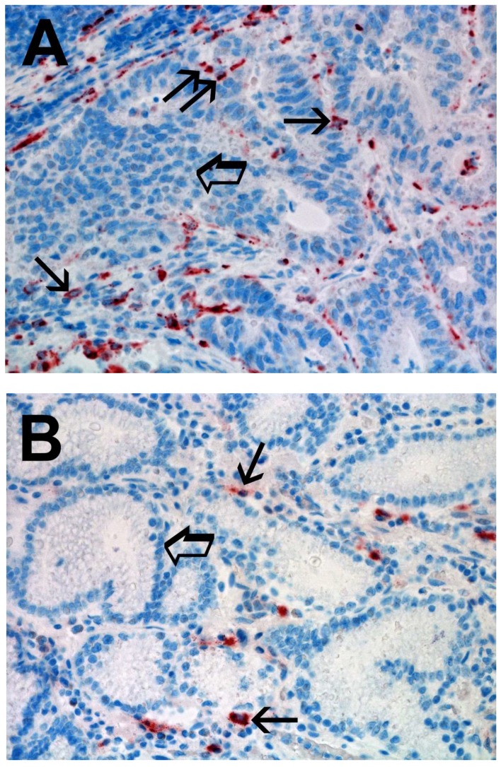
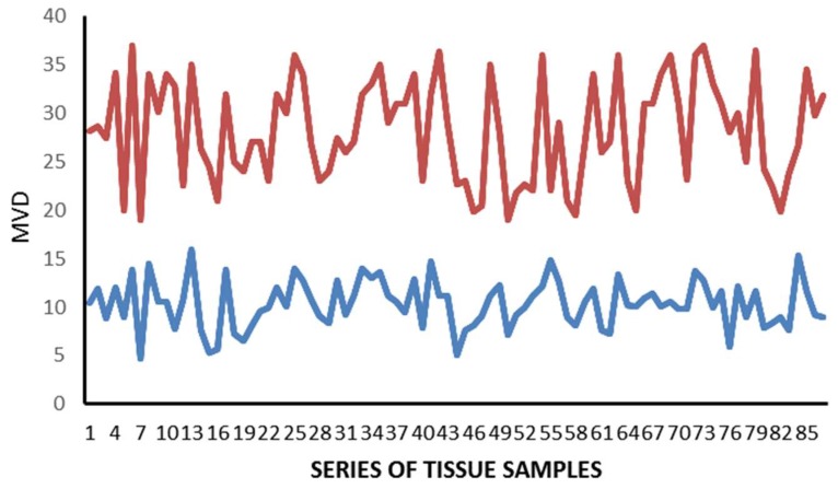
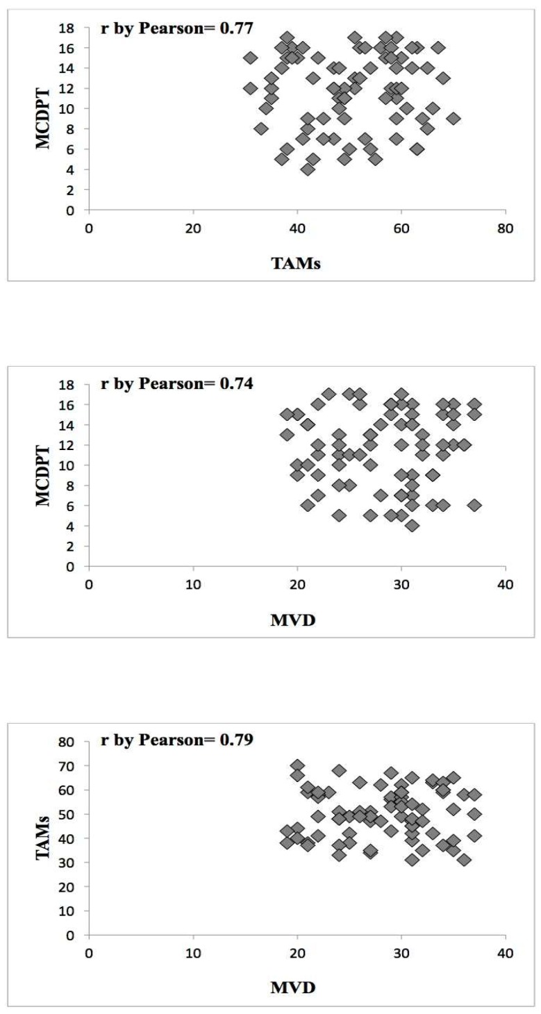
Mast cells and macrophages can play a role in tumor angiogenesis by stimulating microvascular density (MVD). The density of mast cells positive to tryptase (MCDPT), tumor-associated macrophages (TAMs), and MVD were evaluated in a series of 86 gastric cancer (GC) tissue samples from patients who had undergone potential curative surgery. MCDPT, TAMs, and MVD were assessed in tumor tissue (TT) and in adjacent normal tissue (ANT) by immunohistochemistry and image analysis. Each of the above parameters was correlated with the others and, in particular for TT, with important clinico-pathological features. In TT, a significant correlation between MCDPT, TAMs, and MVD was found by Pearson t-test analysis (p ranged from 0.01 to 0.02). No correlation to the clinico-pathological features was found. A significant difference in terms of mean MCDPT, TAMs, and MVD between TT and ANT was found (p ranged from 0.001 to 0.002). Obtained data suggest MCDPT, TAMs, and MVD increased from ANT to TT. Interestingly, MCDPT and TAMs are linked in the tumor microenvironment and they play a role in GC angiogenesis in a synergistic manner. The assessment of the combination of MCDPT and TAMs could represent a surrogate marker of angiogenesis and could be evaluated as a target of novel anti-angiogenic therapies in GC patients.
Keywords: angiogenesis; gastric cancer; macrophages; mast cells; microvascular density; tryptase.
Conflict of interest statement
The authors declare no conflict of interest.
Figures


References
-
- Marech I., Ammendola M., Sacco R., Sammarco G., Zuccalà V., Zizzo N., Leporini C., Luposella M., Patruno R., Filippelli G., et al. Tumour-associated macrophages correlate with microvascular bed extension in colorectal cancer patients. J. Cell. Mol. Med. 2016;20:1373–1380. doi: 10.1111/jcmm.12826. - DOI - PMC - PubMed
-
- Patruno R., Marech I., Zizzo N., Ammendola M., Nardulli P., Gadaleta C., Introna M., Capriuolo G., Rubini R.A., Ribatti D., et al. c-Kit expression, angiogenesis, and grading in canine mast cell tumour: A unique model to study c-Kit driven human malignancies. Biomed. Res. Int. 2014;2014:730246. doi: 10.1155/2014/730246. - DOI - PMC - PubMed
MeSH terms
Substances
LinkOut - more resources
Full Text Sources
Other Literature Sources
Medical
Miscellaneous
