

Preoperative dynamic breast magnetic resonance imaging kinetic features using computer-aided diagnosis: Association with survival outcome and tumor aggressiveness in patients with invasive breast cancer
- PMID: 29649266
- PMCID: PMC5896992
- DOI: 10.1371/journal.pone.0195756
Preoperative dynamic breast magnetic resonance imaging kinetic features using computer-aided diagnosis: Association with survival outcome and tumor aggressiveness in patients with invasive breast cancer
Abstract
Objectives: To evaluate whether preoperative breast dynamic contrast-enhanced (DCE) magnetic resonance (MR) imaging kinetic features, assessed using computer-aided diagnosis (CAD), can predict survival outcome and tumor aggressiveness in patients with invasive breast cancer.
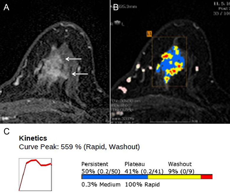
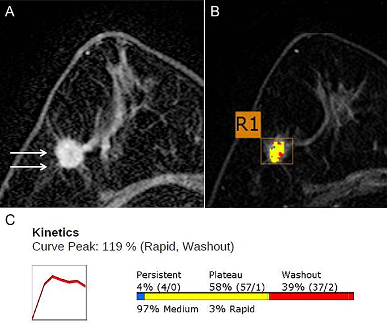
Materials and methods: Between March and December 2011, 301 women who underwent preoperative DCE MR imaging for invasive breast cancer, with CAD data, were identified. All MR images were retrospectively evaluated using a commercially available CAD system. The following kinetic parameters were prospectively recorded for each lesion: initial peak enhancement, the proportion of early phase medium and rapid enhancement, and the proportion of delayed phase persistent, plateau, and washout enhancement. The Cox proportional hazards model was used to determine the association between the kinetic features assessed by CAD and disease-free survival (DFS). The peak signal intensity and kinetic enhancement profiles were compared with the clinical-pathological variables.
Results: There were 32 recurrences during a mean follow-up time of 55.2 months (range, 5-72 months). Multivariate analysis revealed that a higher peak enhancement (DFS hazard ratio, 1.004 [95% confidence interval (CI): 1.001, 1.006]; P = .013) on DCE MR imaging and a triple-negative subtype (DFS hazard ratio, 21.060 [95% CI: 2.675, 165.780]; P = .004) were associated with a poorer DFS. Higher peak enhancement was significantly associated with a higher tumor stage, clinical stage, and histologic grade.
Conclusions: Patients with breast cancer who showed higher CAD-derived peak enhancement on breast MR imaging had worse DFS. Peak enhancement and volumetric analysis of kinetic patterns were useful for predicting tumor aggressiveness.
Conflict of interest statement
Figures
References
-
- Houssami N, Ciatto S, Macaskill P, Lord SJ, Warren RM, Dixon JM, et al. Accuracy and surgical impact of magnetic resonance imaging in breast cancer staging: systematic review and meta-analysis in detection of multifocal and multicentric cancer. J Clin Oncol. 2008;26(19):3248–58. Epub 2008/05/14. doi: 10.1200/JCO.2007.15.2108 . - DOI - PubMed
-
- Warner E, Messersmith H, Causer P, Eisen A, Shumak R, Plewes D. Systematic review: using magnetic resonance imaging to screen women at high risk for breast cancer. Ann Intern Med. 2008;148(9):671–9. Epub 2008/05/07. . - PubMed
-
- Kuhl CK, Mielcareck P, Klaschik S, Leutner C, Wardelmann E, Gieseke J, et al. Dynamic breast MR imaging: are signal intensity time course data useful for differential diagnosis of enhancing lesions? Radiology. 1999;211(1):101–10. Epub 1999/04/06. doi: 10.1148/radiology.211.1.r99ap38101 . - DOI - PubMed
-
- Tuncbilek N, Tokatli F, Altaner S, Sezer A, Ture M, Omurlu IK, et al. Prognostic value DCE-MRI parameters in predicting factor disease free survival and overall survival for breast cancer patients. Eur J Radiol. 2012;81(5):863–7. Epub 2011/03/15. doi: 10.1016/j.ejrad.2011.02.021 . - DOI - PubMed
-
- Turetschek K, Huber S, Floyd E, Helbich T, Roberts TP, Shames DM, et al. MR imaging characterization of microvessels in experimental breast tumors by using a particulate contrast agent with histopathologic correlation. Radiology. 2001;218(2):562–9. Epub 2001/02/13. doi: 10.1148/radiology.218.2.r01fe37562 . - DOI - PubMed
MeSH terms
LinkOut - more resources
Full Text Sources
Other Literature Sources
Medical
Miscellaneous
