

A low dose lipid infusion is sufficient to induce insulin resistance and a pro-inflammatory response in human subjects
- PMID: 29649324
- PMCID: PMC5897027
- DOI: 10.1371/journal.pone.0195810
A low dose lipid infusion is sufficient to induce insulin resistance and a pro-inflammatory response in human subjects
Abstract
Objective: The root cause behind the low-grade inflammatory state seen in insulin resistant (obesity and type 2 diabetes) states is unclear. Insulin resistant subjects have elevations in plasma free fatty acids (FFA), which are ligands for the pro-inflammatory toll-like receptor (TLR)4 pathway. We tested the hypothesis that an experimental elevation in plasma FFA (within physiological levels) in lean individuals would upregulate TLR4 and activate downstream pathways (e.g., MAPK) in circulating monocytes.
Research design and methods: Twelve lean, normal glucose-tolerant subjects received a low dose (30 ml/h) 48 h lipid or saline infusion on two different occasions. Monocyte TLR4 protein level, MAPK phosphorylation, and expression of genes in the TLR pathway were determined before and after each infusion.
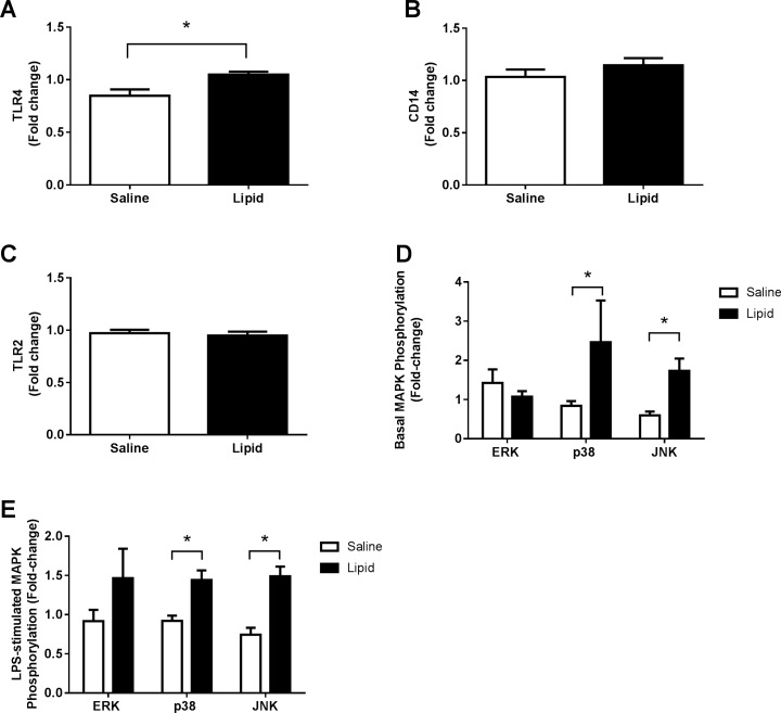
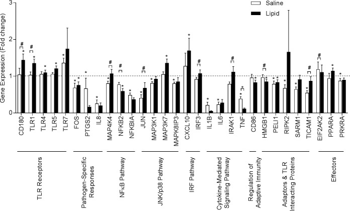
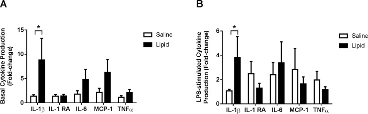
Results: The lipid infusion significantly increased monocyte TLR4 protein and phosphorylation of JNK and p38 MAPK. Lipid-mediated increases in TLR4 and p38 phosphorylation directly correlated with reduced peripheral insulin sensitivity (M value). Lipid increased levels of multiple genes linked to inflammation, including several TLRs, CD180, MAP3K7, and CXCL10. Monocytes exposed in vivo to lipid infusion exhibited enhanced in vitro basal and LPS-stimulated IL-1β secretion.
Conclusions: In lean subjects, a small increase in plasma FFA (as seen in insulin resistant subjects) is sufficient to upregulate TLR4 and stimulate inflammatory pathways (MAPK) in monocytes. Moreover, lipids prime monocytes to endotoxin. We provide proof-of-concept data in humans indicating that the low-grade inflammatory state characteristic of obesity and type 2 diabetes could be caused (at least partially) by pro-inflammatory monocytes activated by excess lipids present in these individuals.
Conflict of interest statement
Figures
References
-
- Hotamisligil GS, Shargill NS, Spiegelman BM. Adipose expression of tumor necrosis factor-alpha: direct role in obesity-linked insulin resistance. Science. 1993. January 1;259(5091):87–91. - PubMed
-
- Weisberg SP, McCann D, Desai M, Rosenbaum M, Leibel RL, Ferrante AW Jr . Obesity is associated with macrophage accumulation in adipose tissue. J Clin Invest. 2003. December;112(12):1796–808. doi: 10.1172/JCI19246 - DOI - PMC - PubMed
-
- Yin MJ, Yamamoto Y, Gaynor RB. The anti-inflammatory agents aspirin and salicylate inhibit the activity of I(kappa)B kinase-beta. Nature. 1998. November 5;396(6706):77–80. doi: 10.1038/23948 - DOI - PubMed
-
- Viardot A, Heilbronn LK, Samocha-Bonet D, Mackay F, Campbell LV, Samaras K. Obesity is associated with activated and insulin resistant immune cells. Diabetes Metab Res Rev. 2012. July;28(5):447–54. doi: 10.1002/dmrr.2302 - DOI - PubMed
-
- van Oostrom AJ, van Wijk JP, Sijmonsma TP, Rabelink TJ, Castro Cabezas M. Increased expression of activation markers on monocytes and neutrophils in type 2 diabetes. Neth J Med. 2004. October;62(9):320–5. - PubMed
Publication types
MeSH terms
Substances
Grants and funding
- P30 AG044271 /DK/NIDDK NIH HHS/United States
- R01-DK80157/DK/NIDDK NIH HHS/United States
- P30 AG013319/AG/NIA NIH HHS/United States
- UL1TR000149 /DK/NIDDK NIH HHS/United States
- R01 DK089229/DK/NIDDK NIH HHS/United States
- R01 DK080157/DK/NIDDK NIH HHS/United States
- UL1 TR000149/TR/NCATS NIH HHS/United States
- P30 AG044271/AG/NIA NIH HHS/United States
- AG013319 /DK/NIDDK NIH HHS/United States
- R01-DK089229/DK/NIDDK NIH HHS/United States
- P30 CA054174/CA/NCI NIH HHS/United States
- UL1 TR001120/TR/NCATS NIH HHS/United States
LinkOut - more resources
Full Text Sources
Other Literature Sources
Research Materials
Miscellaneous
