. 2018 May 20:120:407-413.
doi: 10.1016/j.freeradbiomed.2018.04.009. Epub 2018 Apr 9.

Nrf2 signaling and autophagy are complementary in protecting breast cancer cells during glucose deprivation

Affiliations

Nrf2 signaling and autophagy are complementary in protecting breast cancer cells during glucose deprivation

Alyssa Walker et al. Free Radic Biol Med. .

Abstract

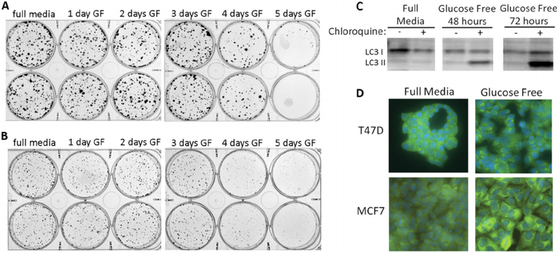
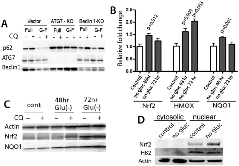
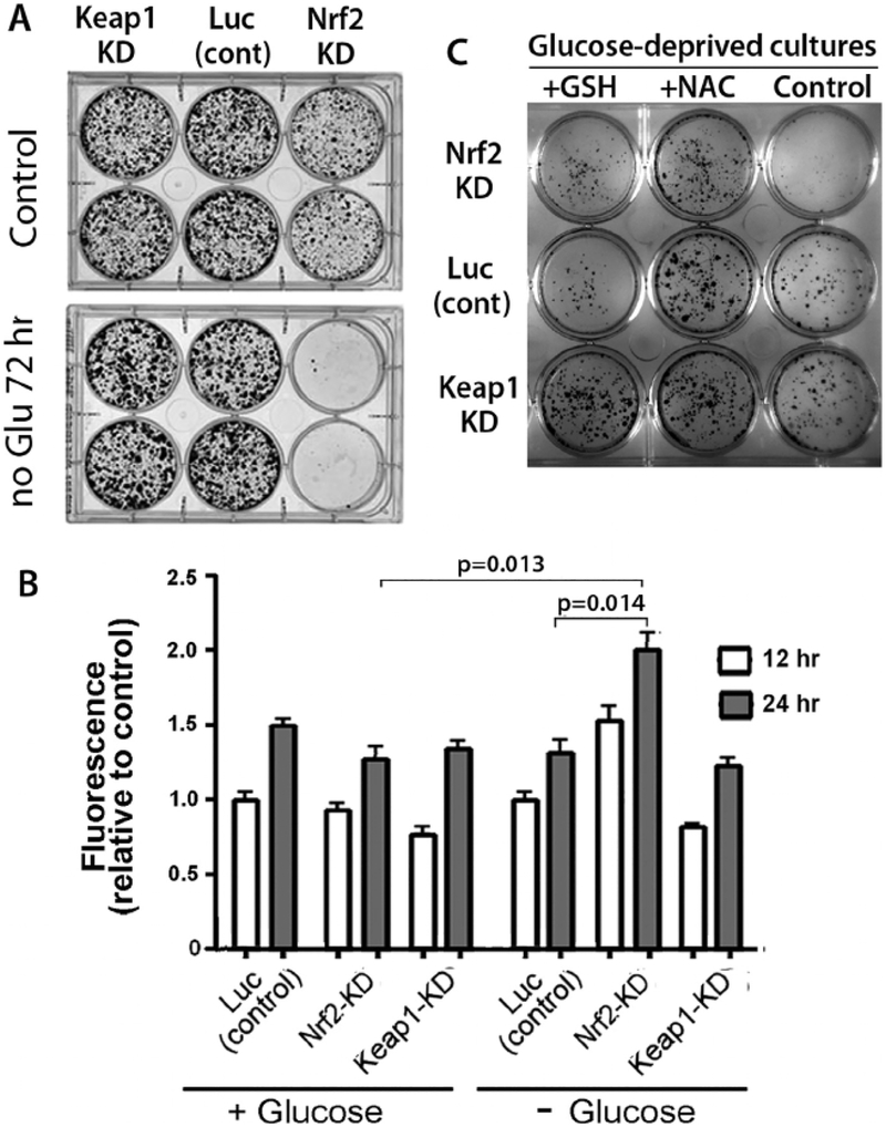
Autophagy can serve as a mechanism for survival of cells during nutrient deprivation by recycling cellular macromolecules and organelles transiently to provide essential metabolic substrates. However, autophagy itself causes metabolic stress to cells, and other cellular protective mechanisms likely cooperate with autophagy to promote cell survival during nutrient deprivation. In this study, we explored protective mechanisms in breast cancer cells in the setting of glucose deprivation. While breast cancer cells (MCF7 and T47D) survive in glucose-free medium for three days or more, autophagy is induced in this setting. Blocking autophagy pharmacologically with chloroquine or by knock-out of an essential autophagy gene, such as Beclin 1 or ATG7, markedly reduces the ability of cells to survive during glucose deprivation. Autophagy previously was shown to degrade p62, a protein that sequesters KEAP1, and KEAP1 in turn sequesters Nrf2, a master regulator of the antioxidant response. Hence, we investigated how the Nrf2 signaling pathway might be affected by glucose deprivation and autophagy. We found that while glucose deprivation does cause decreased cellular levels of p62, Nrf2 protein levels and activity unexpectedly increase in this setting. Moreover, this increase in Nrf2 activity provides important protection to breast cancer cells during glucose deprivation, since siRNA knockdown of Nrf2 markedly impairs survival during glucose deprivation. Antioxidants, N-acetyl cysteine and glutathione also protect these cells during glucose deprivation, leading us to conclude that Nrf2 signaling via its antioxidant activity has a critical and previously undescribed role of protecting cells during glucose deprivation-induced autophagy.

Keywords: ATG7; Antioxidant response; Autophagy; Beclin-1; KEAP1; Nrf2; P62.


Figures

Fig. 1.
Fig. 1.. Glucose deprivation causes autophagy in breast cancer cells.
Breast cancer cells (MCF7, panel A; T47D, panel B) plated at clonal density were cultured in glucose-free medium for up to 5 days, and then allowed to form colonies in full medium for an additional 8–10 days. Panel C: glucose deprivation in MCF7 cells causes increases in LC3 II to LC3I ratio, particularly when completion of autophagy is blocked by chloroquine. Panel D: Glucose deprivation in T47D and MCF7 cells results in appearance of punctate foci of LC3, typical of autophagosomes in macroautophagy.
Fig. 2.
Fig. 2.. Autophagy is protective of cell survival during glucose deprivation.
Panel A shows effects of chloroquine, which blocks completion of autophagy, on survival of MCF7 cells cultured with full media or glucose-free medium for 3 days, followed by full medium for 10 days. In panel B, cultures grown in glucose-free medium are seen to fragment in the presence of chloroquine, suggesting that apoptosis is induced when autophagy is blocked. Panel C confirms that poly-ADP ribose polymerase is cleaved in glucose-deprived cells cultured with chloroquine. Panel D shows that, with CRISPR/ Cas9 knock-out of ATG7 or Beclin 1, MCF7 cells have markedly reduced survival after glucose deprivation for 3 days.
Fig. 3.
Fig. 3.. Glucose deprivation leads to decreased levels of p62 and increased levels of Nrf2 in MCF7 breast cancer cells.
Panel A shows decreased levels of p62 in MCF7 cells cultured for 72 h in glucose-free medium (G-F). These decreases in p62 are dependent on autophagy and do not occur in cells with knock-out of Belcin 1 or ATG7. Panel B: Time dependent changes in transcripts for Nrf2 and its target genes, heme oxygenase 1 (HMOX) and nitroquinone oxidase 1 (NQO1), during glucose deprivation. Fold changes were calculated relative to control cultures in complete media, and -actin was used for as endogenous control for each measurement. Measurements from glucose-deprived cultures were then compared to respective control cultures by t-test, with p values shown. Panel C: Immunoblots show that protein levels of both Nrf2 and NQO1 increase after 72 h glucose deprivation, and Panel D shows that the increased levels of Nrf2 protein are predominantly within the nuclear fraction of fractionated cells.
Fig. 4.
Fig. 4.. Nrf2 provides antioxidant protection of breast cancer cells during glucose deprivation.
Panel A demonstrates that survival of MCF7 cells during glucose deprivation is dependent on Nrf2, since knock-down of Nrf2 leads to markedly decreased survival in cells cultured in glucose-deficient medium for 3 days. Panel B shows measurements of reactive oxygen species using the fluorescent marker, DCFDA. Note the significantly greater levels of ROS in cells that had knock-down of Nrf2 compared to cells with knock-down of KEAP1, consistent with Nrf2 activity controlling levels of ROS during glucose deprivation. In panel C, cultures of cells with KEAP1 knock-down, Nrf2 knock-down, or luciferase siRNA control transfection were cultured for 72 h in glucose free medium, followed by growth in full medium for 10 days. Addition of 2.0 mM N-acetyl cysteine (NAC) or 2.5 mM glutathione (GSH) to medium for the full 72 h resulted in improved survival of cultured cells.

References

    1. Rabinowitz JD, White E, Autophagy and metabolism, Science 330 (6009) (2010) 1344–1348, 10.1126/science.1193497. - DOI - PMC - PubMed
    1. Moruno F, Perez-Jimenez E, Knecht E, Regulation of autophagy by glucose in Mammalian cells, Cells 1 (3) (2012) 372–395, 10.3390/cells1030372. - DOI - PMC - PubMed
    1. Xi H, Kurtoglu M, Liu H, Wangpaichitr M, You M, Liu X, Savaraj N, Lampidis TJ, 2-Deoxy-D-glucose activates autophagy via endoplasmic reticulum stress rather than ATP depletion, Cancer Chemother. Pharmacol 67 (4) (2011) 899–910, 10.1007/s00280-010-1391-0. - DOI - PMC - PubMed
    1. Altman BJ, Jacobs SR, Mason EF, Michalek RD, MacIntyre AN, Coloff JL, Ilkayeva O, Jia W, He YW, Rathmell JC, Autophagy is essential to suppress cell stress and to allow BCR-Abl-mediated leukemogenesis, Oncogene 30 (16) (2011) 1855–1867, 10.1038/onc.2010.561. - DOI - PMC - PubMed
    1. Jeon JY, Kim SW, Park KC, Yun M, The bifunctional autophagic flux by 2-deoxyglucose to control survival or growth of prostate cancer cells, BMC Cancer 15 (2015) 623, 10.1186/s12885-015-1640-z. - DOI - PMC - PubMed