Comparative Study
. 2018 Jun:5:172-179.
doi: 10.1016/j.pvr.2018.04.002. Epub 2018 Apr 9.

HPV DNA methylation at the early promoter and E1/E2 integrity: A comparison between HPV16, HPV18 and HPV45 in cervical cancer

Affiliations
Comparative Study

HPV DNA methylation at the early promoter and E1/E2 integrity: A comparison between HPV16, HPV18 and HPV45 in cervical cancer

Sérgio Menezes Amaro-Filho et al. Papillomavirus Res. 2018 Jun.

Abstract

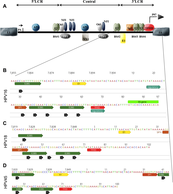
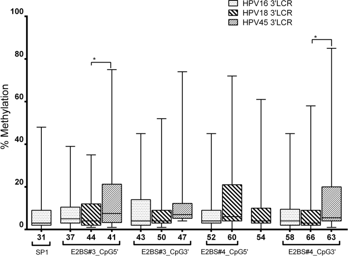
Objectives: To compare and describe type-specific characteristics of HPV16, HPV18 and HPV45 in cervical cancer with respect to 3'LCR methylation and disruption of E1/E2.

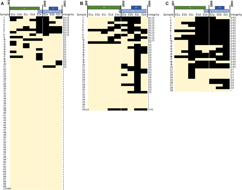
Methods: The methylation level of 137 cervical cancer samples (70 with HPV16, 37 with HPV18, and 30 with HPV45) of Brazilian patients was analyzed by pyrosequencing. PCR amplifications were performed to characterize E1 and E2 disruption as an episomal surrogate.

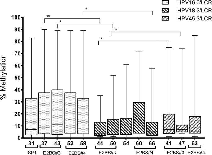
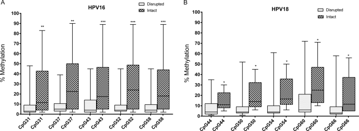
Results: The 3'LCR of HPV16 showed a higher methylation at all CpG sites (7%, 9%, 11%, 10% and 10%) than homologous HPV18 regions (4%, 5%. 6%, 9% and 5%) and HPV45 regions (7%, 7% and 5%). Presence of intact E1/E2 was associated with higher HPV16 and HPV18 methylation levels at all CpG sites (p < 0.05). Disruption of E1/E2 was more frequently found in HPV45 (97%) and HPV18 (84%) than in HPV16 DNA (30%). HPV16 disruption was more frequently found in E1 (48%) unlike HPV18, where it was found in E2 (61%). Concomitant disruption of E1/E2 was most frequent in HPV45 (72%).

Conclusions: The findings showed a higher methylation associated with intact E1/E2 for HPV16 and HPV18. The closely phylogenetic related HPV18 and HPV45 share a similar methylation level and the frequency of viral genome disruption.

Keywords: HPV genome; Human papillomavirus; Invasive cervical cancer; Methylation; Pyrosequencing; Viral genome integration.


Figures

Fig. 1
Fig. 1
Schematic representation of HPV LCR. A, represents the three segments of HPV16 LCR (5′LCR, central or enhancer, and 3′LCR), considered a model of LCR for all HPVs. The 5′LCR contains the transcription termination signal, denoted ‘pA’. The central segment contains the majority of transcription factor binding sites. The 3′LCR contains the origin of replication and the E6/E7 promoter. B, C and D, represent the nucleotide sequence of 3′LCR of HPV16, HPV18 and HPV45, respectively, highlighting the binding motifs of E2, E1, Sp1, TFIID and all CpG sites within this segment. The Sp1 motif of HPV16 contains one CpG site in its core (B, GGGCGT). Differently no CpG is found in the Sp1 motif of HPV18 (C, GGGAGT) and HPV45 (D, GGGTGT). The E2BS#4 of HPV45 (D) has only one CpG site (nt 63), while HPV16 (B) and HPV18 (C) contain two CpG sites. (For information on motifs and reference genome data see references , , and Papillomavirus Episteme database , respectively). All transcription factor binding sites are denoted by the abbreviation used in the text except for TEF-1 that is herein denoted as TF1.
Fig. 2
Fig. 2
Methylation status of homologous CpG sites in the 3′LCR of HPV16, HPV18 and HPV45. The methylation level of each CpG site is represented by a box displaying upper and lower quartiles separated by the median line, and whisker plots. HPV16 displayed a higher level of methylation than HPV18 and HPV45, mainly at E2 binding sites (E2BS#3 and #4). This was statistically different when comparing the homologous CpG sites of HPV16 vs HPV18 (CpG 37 vs 44, p = 0.0018; CpG 43 vs 50, p = 0.0490; and 58 vs 66, p = 0.0109), and HPV18 vs HPV45 (CpG 44 vs CpG41). Analyses were performed with the Mann-Whitney U test.
Fig. 3
Fig. 3
Map of disruptions sites inE1andE2of the 3′LCR of HPV per sample. A, E1 and E2 of HPV16 predominately showed intact genes (light orange color - I), with high disruption of E1. B, integrity of HPV18 E1 and E2, with most samples displaying disrupted DNA, predominately of E2. C displays E1 and E2 integrity of HPV45 showing significant loss of both genes and larger deletions. Black rectangles represent lack of PCR amplification; light orange indicate presence of amplification. D1/2 indicates E1 and E2 disruption; D1: exclusive E1 disruption; D2: exclusive E2 disruption; I: intact E1 and E2. E1a, E1b, E1c, E1d and E1e represent amplicons covering the E1 gene. E2a, E2b, E2c and E2d represent amplicons covering the E2 gene. The genome position of each amplification is described in Table 2.
Fig. 4
Fig. 4
3′LCR of HPV16 and HPV18 respective to E1 and E2 integrity. The methylation level of each CpG site is represented by a box displaying upper and lower quartiles separated by the median line, and whisker plots. A higher methylation at all CpG sites was found in samples with intact E1 and E2 than with disrupted genes (*p < 0.05; **p < 0.005 and ***p < 0.0005, Mann-Whitney Test).
Fig. S1
Fig. S1
Comparison of the methylation between homologous CpG sites at the 3′LCR of HPV in disrupted samples. A higher methylation in E2BS#3 and E2BS#4 of HPV45 (nt 41 and 63) was observed than in HPV18 (nt 44 and 66, respectively); p = 0.0163 and p = 0.0246, respectively. Homologous CpG sites in E2 binding site motifs of each HPV genotype comprised: (i) nt 37, 44 and 41 of HPV16, HPV18 and HPV45, respectively, at the 5′ end of E2BS#3; (ii) nt 43, 50 and 47 of HPV16, HPV18 and HPV45, respectively, at the 3′ end of E2BS#3; (iii) nt 52 and 60 of HPV16 and HPV18, respectively, at the 5′ end of E2BS#4; and (iv) nt 58, 66 and 63 of HPV16, HPV18 and HPV45, respectively, at the 3′ end of E2BS#4. Associations approaching borderline statistical significance: nt 47 vs 43 (p = 0.0668), nt 60 vs 52 (p = 0.0995) and 63 vs 58 (p = 0.0716).

References

    1. Doorbar J. Molecular biology of human papillomavirus infection and cervical cancer. Clin. Sci. 2006;110:525–541. - PubMed
    1. IARC Working group on the evaluation of carcinogenic risks to human. biological agents. A review of human carcinogens. IARC Monogr. Eval. Carcinog. Risks Hum. 2012;100B:1–441. - PMC - PubMed
    1. Mark O′Connor S.-Y.C., Bernard Hans-Ulrich. Transcription Factor Binding Sites in the Long Control Region of Genital HPVs. In: Myers G., Bernard H.U., Delius H., Baker K., Icenogle J., Halpern A., Wheeler C., editors. Human Papillomaviruses, 1995 Compendium, Part III. Los Alamos National Laboratory; Los Alamos, NMex: 1995. pp. 21–40.
    1. Tan S.H., Bartsch D., Schwarz E., Bernard H.U. Nuclear matrix attachment regions of human papillomavirus type 16 point toward conservation of these genomic elements in all genital papillomaviruses. J. Virol. 1998;72:3610–3622. - PMC - PubMed
    1. Bernard H.U. Gene expression of genital human papillomaviruses and considerations on potential antiviral approaches. Antivir. Ther. 2002;7:219–237. - PubMed

Publication types

Substances