. 2018 Jul:170:26-36.
doi: 10.1016/j.biomaterials.2018.03.054. Epub 2018 Mar 31.

Polymeric micelles: Theranostic co-delivery system for poorly water-soluble drugs and contrast agents

Affiliations

Polymeric micelles: Theranostic co-delivery system for poorly water-soluble drugs and contrast agents

Jaydev R Upponi et al. Biomaterials. 2018 Jul.

Abstract

Interest in theranostic agents has continued to grow because of their promise for simultaneous cancer detection and therapy. A platform-based nanosized combination agent suitable for the enhanced diagnosis and treatment of cancer was prepared using polymeric polyethylene glycol-phosphatidylethanolamine-based micelles loaded with both, poorly soluble chemotherapeutic agent paclitaxel and hydrophobic superparamagnetic iron oxide nanoparticles (SPION), a Magnetic Resonance Imaging contrast agent. The co-loaded paclitaxel and SPION did not affect each other's functional properties in vitro. In vivo, the resulting paclitaxel-SPION-co-loaded PEG-PE micelles retained their Magnetic Resonance contrast properties and apoptotic activity in breast and melanoma tumor mouse models. Such theranostic systems are likely to play a significant role in the combined diagnosis and therapy that leads to a more personalized and effective form of treatment.

Keywords: Cancer therapy; Diagnostics; Drug delivery; Micelles; Nanoparticles; Theranostics.


Figures

Figure 1
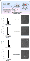
Figure 1. Characterization of micellar theranostic agent
Scheme (A) illustrates a PEG-PE micellar theranostic agent co-incorporated with paclitaxel and MRI contrast agents along with the chemical structure of PEG-PE showing the hydrophilic PEG which depicts the micelle corona and core-hydrophobic PE group, responsible for the solubilization of PTX and entrapment of SPIONs. (B) indicates micelle size and size distribution of empty PEG-PE/Plain micelles, PTX-loaded PEG-PE micelles, SPION-loaded PEG-PE micelles and co-loaded PTX/SPION PEG-PE theranostic micelles with their respective electron micrographs (C) at 75,000x magnification, scale bar repesents 100nm. (n=3)
Figure 2
Figure 2. T2-weighted MR image and relaxation times
MR imaging of contrast agent in the absence (A) and presence (B) of 1.5% w/w PTX during formulation optimization and their T2 relaxation times (D). T2-weighted MR images of 3% SPION-containing micelles and controls (C) along with their respective T2 relaxation times (E). MR images were obtained using a 7T Bruker magnet. T2 relaxation times were calculated using Paravision 5.0 software. (mean ± S.D., n=3).
Figure 3
Figure 3. In vitro cytotoxicity of micellar formulations
Percent viability of 4T1 and B16F10 cells after treatment with plain, SPION, PTX, theranostic micelles or free drug (PTX) at +24 hours (A, B) and + 48 hours (C, D) incubation. Data represent mean ± S.D. (n=3). (*) p < 0.05 between paclitaxel micelles and free paclitaxel; (#) p < 0.05 between theranostic micelles and free paclitaxel (Student’s t-test).
Figure 4
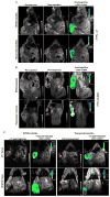
Figure 4. Whole body in vivo images
T-2 weighted MR image and color-coded composite maps acquired with breast (A) and melanoma (B) tumor-bearing mice, pre- and post-injection of various formulations containing SPIONs as MR contrast agent. Images were obtained using a 7 Tesla Bruker scanner. Similar images (C) were acquired 72 hours post-injection from both tumor models. Non-color coded images show regions of the tumor bounded by a dotted line. The arrow points the accumulation of contrast agent at the tumor site. Theranostic micelles retained their MR contrast properties equivalent to that of SPION-only micelles.
Figure 5
Figure 5. Prussian blue staining of tumor cryosections
Breast (4T1) and melanoma (B16F10) tumor sections stained for SPIONs localized within the tumor interstitium at +48 (A) and +72 hours (B). Blue stain (as indicated by the arrow) from the sections indicates the presence of SPION micelles against a light-red to pink background counterstained with nuclear red that stains cytoplasm and nucleus. The melanoma cells are characterized by their natural dark brown pigments. (Scale bar = 1cm)
Figure 6
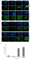
Figure 6. TUNEL assay for apoptotic activity of paclitaxel
In vivo tumor cell apoptosis was induced by intra-tumoral administration of PTX-containing formulations in breast (4T1) and melanoma (B16F10) tumor-bearing mice. At +48 hours (A, B) and +72 hours (C, D) tumor apoptosis was observed after treatment with theranostic and PTX-only-containing formulations as determined by TUNEL assay. Panel (E) shows bar graph of average fluorescence intensity in arbitrary units across all formulations, (n=5) and data is represented as mean±S.D. Cells undergoing apoptosis were observed in the green/FITC channel (see arrow). Sections observed with a blue filter indicate DAPI stain for nuclei. In both cases, tumors treated with plain and SPION micelles lacked DNA fragmenting cells. (Scale bar = 1cm).

Similar articles

Cited by

References

    1. Rai P, Mallidi S, Zheng X, Rahmanzadeh R, Mir Y, Elrington S, Khurshid A, Hasan T. Development and applications of photo-triggered theranostic agents. Adv Drug Deliv Rev. 2010;62(11):1094–124. - PMC - PubMed
    1. MacKay JA, Li Z. Theranostic agents that co-deliver therapeutic and imaging agents? Adv Drug Deliv Rev. 2010;62(11):1003–4. - PubMed
    1. Maeda H, Wu J, Sawa T, Matsumura Y, Hori K. Tumor vascular permeability and the EPR effect in macromolecular therapeutics: a review. J Control Release. 2000;65(1–2):271–84. - PubMed
    1. Khemtong C, Kessinger CW, Gao J. Polymeric nanomedicine for cancer MR imaging and drug delivery. Chem Commun. 2009;(24):3497–510. - PMC - PubMed
    1. Hahn MA, Singh AK, Sharma P, Brown SC, Moudgil BM. Nanoparticles as contrast agents for in-vivo bioimaging: current status and future perspectives. Anal Bioanal Chem. 2011;399(1):3–27. - PubMed

Publication types

MeSH terms

LinkOut - more resources