Comparative Study
. 2018 Jul 31;19(4):483-491.
doi: 10.4142/jvs.2018.19.4.483.

Comparison of electrophysiological properties of two types of pre-sympathetic neurons intermingled in the hypothalamic paraventricular nucleus

Affiliations
Comparative Study

Comparison of electrophysiological properties of two types of pre-sympathetic neurons intermingled in the hypothalamic paraventricular nucleus

Yiming Shen et al. J Vet Sci. .

Abstract

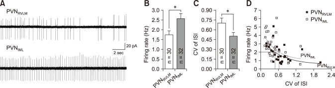
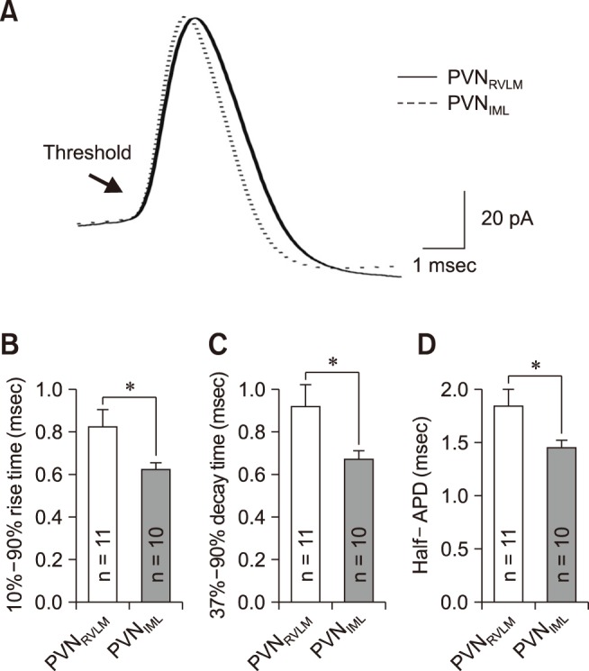
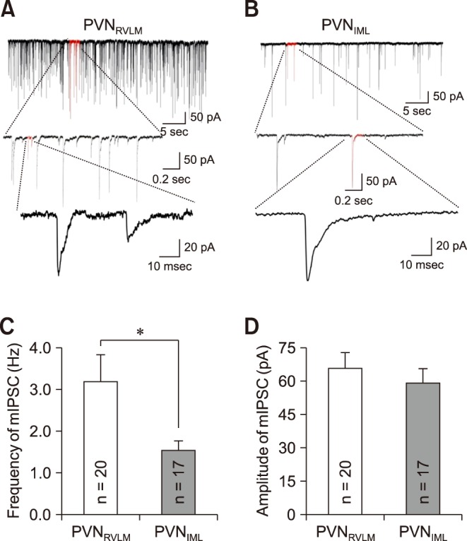
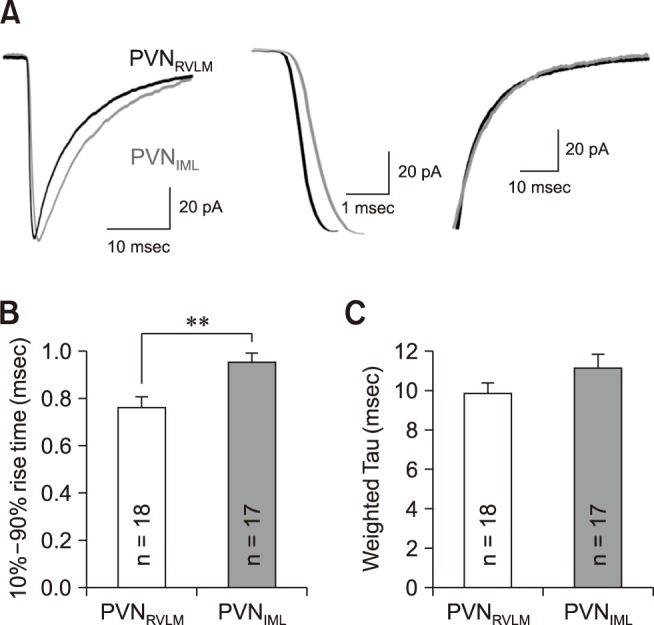
The hypothalamic paraventricular nucleus (PVN) contains two types of neurons projecting to either the rostral ventrolateral medulla (PVNRVLM) or the intermediolateral horn (IML) of the spinal cord (PVNIML). These two neuron groups are intermingled in the same subdivisions of the PVN and differentially regulate sympathetic outflow. However, electrophysiological evidence supporting such functional differences is largely lacking. Herein, we compared the electrophysiological properties of these neurons by using patch-clamp and retrograde-tracing techniques. Most neurons (>70%) in both groups spontaneously fired in the cell-attached mode. When compared to the PVNIML neurons, the PVNRVLM neurons had a lower firing rate and a more irregular firing pattern (p < 0.05). The PVNRVLM neurons showed smaller resting membrane potential, slower rise and decay times, and greater duration of spontaneous action potentials (p < 0.05). The PVNRVLM neurons received greater inhibitory synaptic inputs (frequency, p < 0.05) with a shorter rise time (p < 0.05). Taken together, the results indicate that the two pre-sympathetic neurons differ in their intrinsic and extrinsic electrophysiological properties, which may explain the lower firing activity of the PVNRVLM neurons. The greater inhibitory synaptic inputs to the PVNRVLM neurons also imply that these neurons have more integrative roles in regulation of sympathetic activity.

Keywords: action potential; inhibitory postsynaptic current; patch-clamp techniques; rostral ventrolateral medulla; spinal cord lateral horn.


Conflict of interest statement

Conflict of Interest: The authors declare no conflicts of interest.

Figures

Fig. 1
Fig. 1. The firing activity in PVNRVLM and PVNIML neurons. (A) Representative traces showing spontaneous firing activities of a PVNRVLM neuron and a PVNIML neuron. (B and C) Cumulative bar graphs showing the mean frequency of spontaneous spikes (B) and the coefficient of variance (CV) of the interspike interval (ISI) in PVNRVLM and PVNIML neurons (C). (D) Plot of firing rate vs. the CV of the ISI. The curves were drawn by fitting the data with the equation y = a + b × cx. The CV values in the PVNIML neurons tended to be smaller than those in the PVNRVLM neurons at the same firing rate. The values in the bars (B and C) represent the total number of neurons tested. Values represent mean ± SEM. Asterisks indicate p < 0.05 obtained by the unpaired Student's t-test. PVN, paraventricular nucleus; RVLM, rostral ventrolateral medulla; IML, intermediolateral horn of the spinal cord.
Fig. 2
Fig. 2. The parameters of spontaneous action potentials in PVNRVLM and PVNIML neurons. (A) Representative action potentials from a PVNRVLM neuron (solid line; RMP, −56.07 mV; threshold, −43.09 mV; rise time, 0.95 msec; decay time, 1.05 msec; half-APD, 2.15 msec) and a PVNIML neuron (dotted line; RMP, −60.53 mV; threshold, −39.93 mV; rise time, 0.6 msec; decay time, 0.75 msec; half-APD, 1.65 msec) were overlapped at threshold. Cumulative bar graphs showing the 10% to 90% rise time (B), 37% to 90% decay time (C) and half-APD (D) in PVNRVLM and PVNIML neurons. The values in the bars (B–D) represent the total number of neurons tested. Values represent mean ± SEM. Asterisks indicate p < 0.05 by the unpaired Student's t-test. PVN, paraventricular nucleus; RVLM, rostral ventrolateral medulla; IML, intermediolateral horn of the spinal cord; half-APD, duration of action potential at half amplitude.
Fig. 3
Fig. 3. Miniature inhibitory postsynaptic current (mIPSC) in PVNRVLM and PVNIML neurons. Representative current traces of mIPSCs in a PVNRVLM neuron (A) and a PVNIML neuron (B) (Vh = −70 mV). Cumulative bar graphs showing the mean frequency (C) and mean amplitude (D) of the mIPSCs. The values in the bars (C and D) represent the total numbers of neurons tested. Values are mean ± SEM. Asterisk indicates p < 0.05 by the unpaired Student's t-test. PVN, paraventricular nucleus; RVLM, rostral ventrolateral medulla; IML, intermediolateral horn of the spinal cord.
Fig. 4
Fig. 4. The miniature inhibitory postsynaptic current (mIPSC) kinetics in the PVNRVLM and the PVNIML neurons. (A) Representative traces of mIPSCs in a PVNRVLM neuron (black) and a PVNIML neuron (gray) (left). Illustration of the 10% to 90% rise time and the decay time at an expanded time scale (middle and right). (B) Summary bar graphs showing the mean 10% to 90% rise time in the PVNRVLM and PVNIML neurons. (C) Summary bar graphs displaying the weighted decay time constant under a well-fitted second exponential. The values in the bars (B and C) represent the total numbers of neurons tested. Values are mean ± SEM. Double asterisk indicates p < 0.05 by the unpaired Student's t-test. PVN, paraventricular nucleus; RVLM, rostral ventrolateral medulla; IML, intermediolateral horn of the spinal cord.

References

    1. Abbott LF, Fusi S, Miller KD. Theoretical approaches to neuroscience: examples from single neurons to networks. In: Kandel ER, Schwartz JH, Jessell TM, Siegelbaum SA, Hudspeth AJ, Mack S, editors. Principles of Neural Science. 5th ed. New York: McGraw-Hill Professional Publishing; 2012. pp. 1601–1617.
    1. Badoer E. Hypothalamic paraventricular nucleus and cardiovascular regulation. Clin Exp Pharmacol Physiol. 2001;28:95–99. - PubMed
    1. Berkefeld H, Fakler B. Repolarizing responses of BKCa-Cav complexes are distinctly shaped by their Cav subunits. J Neurosci. 2008;28:8238–8245. - PMC - PubMed
    1. Brown CH, Bains JS, Ludwig M, Stern JE. Physiological regulation of magnocellular neurosecretory cell activity: integration of intrinsic, local and afferent mechanisms. J Neuroendocrinol. 2013;25:678–710. - PMC - PubMed
    1. Carrive P, Gorissen M. Premotor sympathetic neurons of conditioned fear in the rat. Eur J Neurosci. 2008;28:428–446. - PubMed

Publication types