

A rare presentation of pulmonary sarcoidosis as a solitary lung mass: a case report
- PMID: 29650028
- PMCID: PMC5897926
- DOI: 10.1186/s13256-018-1632-0
A rare presentation of pulmonary sarcoidosis as a solitary lung mass: a case report
Abstract
Background: Sarcoidosis is a multisystem, chronic granulomatous disease of unknown etiology that predominantly affects the lungs. Pulmonary sarcoidosis classically presents with constitutional symptoms and computed tomographic scan findings of bilateral, symmetric micronodules in a peribronchovascular distribution with upper and middle lung zone predominance accompanied by bilateral, symmetric hilar lymphadenopathy. A solitary lung mass is a rare finding for pulmonary sarcoidosis, and with its associated constitutional symptoms, it strongly mimics a malignancy. We aimed to provide further insight into the broad differential diagnosis of a lung mass by describing our experiences in the care of a patient who presented with clinical and radiographic features of lung cancer who was ultimately found to have an atypical manifestation of stage II pulmonary sarcoidosis.
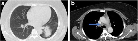
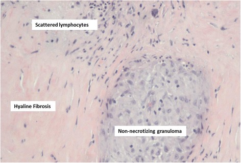
Case presentation: A 44-year-old African American woman with a history of childhood asthma and type 2 diabetes mellitus presented with shortness of breath. After being treated for a presumed asthma exacerbation with prednisone, she experienced worsening dyspnea, night sweats, and unintentional weight loss. Further evaluation revealed a large left lower lobe mass and hilar lymphadenopathy. A computed tomography-guided biopsy of the lung mass revealed a multifocal non-necrotizing granuloma with multinucleated giant cells. Although consistent with sarcoidosis, this finding could represent a sarcoid-like reaction secondary to an occult malignancy. A more extensive repeat biopsy via bronchoscopy and mediastinoscopy revealed granulomatous inflammation without evidence of malignancy or infection. These procedures confirmed the diagnosis of pulmonary sarcoidosis, and she was started on treatment with high-dose prednisone. Her treatment course was complicated by hyperglycemia necessitating insulin therapy, but after 3 months of therapy, she reported improvement in her dyspnea, and repeat imaging revealed a significant decrease in the size of the lung mass and lymphadenopathy. Given her clinical and radiographic response, she was continued on a prednisone taper.
Conclusions: Atypical manifestations of pulmonary sarcoidosis are diagnostically challenging because the clinical and radiographic features of the disease mimic those of a malignancy. We aimed to illustrate a unique etiology of a lung mass and the importance of maintaining a broad differential diagnosis. Nonetheless, with the possibility of a malignancy, a high index of suspicion is necessary for timely diagnosis and optimal management.
Keywords: Lung mass; Pulmonary sarcoidosis; Sarcoid-like reaction.
Conflict of interest statement
Ethics approval and consent to participate
Not applicable.
Consent for publication
Written informed consent was obtained from the patient for publication of this case report and any accompanying images. A copy of the written consent is available for review by the Editor-in-Chief of this journal.
Competing interests
The authors declare that they have no competing interests.
Publisher’s Note
Springer Nature remains neutral with regard to jurisdictional claims in published maps and institutional affiliations.
Figures

References
Publication types
MeSH terms
Substances
Grants and funding
LinkOut - more resources
Full Text Sources
Other Literature Sources
