. 2018 Apr 13;360(6385):204-208.
doi: 10.1126/science.aar3799.

Tropism for tuft cells determines immune promotion of norovirus pathogenesis

Affiliations

Tropism for tuft cells determines immune promotion of norovirus pathogenesis

Craig B Wilen et al. Science. .

Abstract

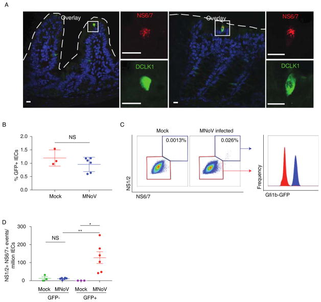
Complex interactions between host immunity and the microbiome regulate norovirus infection. However, the mechanism of host immune promotion of enteric virus infection remains obscure. The cellular tropism of noroviruses is also unknown. Recently, we identified CD300lf as a murine norovirus (MNoV) receptor. In this study, we have shown that tuft cells, a rare type of intestinal epithelial cell, express CD300lf and are the target cell for MNoV in the mouse intestine. We found that type 2 cytokines, which induce tuft cell proliferation, promote MNoV infection in vivo. These cytokines can replace the effect of commensal microbiota in promoting virus infection. Our work thus provides insight into how the immune system and microbes can coordinately promote enteric viral infection.


Figures

Fig. 1
Fig. 1. Fecal-oral MNoV transmission requires radiation-resistant cells
Reciprocal bone marrow transplants were performed among Cd300lf+/+ (WT) and Cd300lf−/− (KO) littermates. Mice were then challenged perorally with MNoVCR6, which establishes persistent enteric infection in WT animals. (A) WT mice remained susceptible to MNoV as measured by viral genomes in feces at indicated time points. In contrast KO mice did not shed MNoVCR6 whether they received WT or KO bone marrow. (B–E) 21 days post-challenge MNoV viral genomes were determined in the ileum (B), colon (C), spleen (D), and mesenteric lymph nodes (MLN) (E). WT recipients had significantly more viral genomes than KO recipients. There was no significant difference between WT recipients of either WT or KO bone marrow. Fecal samples were analyzed by repeated-measures ANOVA. Tissue samples were analyzed by one-way ANOVA. Significant differences for both fecal and tissue samples were compared to WT→WT control as indicated. Shown are means ± SEM. NS, not significant; *P<0.05; **P<0.01; ***P<0.001; ****P<0.0001. L.O.D., limit of detection. Data is pooled from three independent experiments. The number of mice per group is indicated in (B).
Fig. 2
Fig. 2. CD300lf is expressed on tuft cells but not other intestinal epithelial cells
(A–B) The MNoV receptor CD300lf is detectable on rare intestinal epithelial cells with morphology consistent with tuft cells. CD300lf colocalizes with tuft cell markers (A) DCLK1 and (B) CK18 in mouse ileum and colon. CD300lf is apically polarized towards the intestinal lumen. (C) CD300lf is expressed on Gfi1b-GFP+ tuft cells, but not other intestinal epithelial cells as measured by flow cytometry. Events shown are Singlets+Live+CD45EpCAM+. Images and FACS plots are representative of one of at least three independent experiments. Dashed lines represent the epithelial barrier. White boxes in the overlaid image reflect the magnified inset images. Scale bars, 10 microns.
Fig 3
Fig 3. MNoVCR6 specifically infects CD300lf-expressing intestinal tuft cells
(A) MNoV non-structural protein NS6/7 colocalizes with DCLK1 in the ileum and colon of WT mice infected with MNoVCR6 at seven days post-infection. NS6/7 expression is punctate and cytoplasmic consistent with the viral replication complex. (B) Flow cytometry of intestinal epithelial cells (Singlet+Live+CD45EpCAM+) from Gfi1b-GFP+ tuft cell reporter mice reveal similar frequencies of tuft cells between infected and uninfected mice. (C) A rare population of cells that co- express the MNoV non-structural proteins NS1/2 and NS6/7 was observed. These MNoV-positive cells are Gfi1b-GFP+ demonstrating they are tuft cells. (D) NS1/2+NS6/7+ events were significantly enriched among GFP+ cells. NS1/2+NS6/7+ events were at background levels among non-tuft cells. Data is pooled from three independent experiments with one to two mice per group. Shown are means ± SEM. NS, not significant; *P<0.05; **P<0.01. Dashed lines represent the epithelial barrier. White boxes in the overlaid image reflect the magnified inset images. Scale bars, 10 microns.
Fig. 4
Fig. 4. Tuft cell tropism determines transkingdom interactions of MNoV
(A) WT mice were injected intraperitoneally with PBS, IL-4, or IL-25 prior to peroral challenge with a low dose (4.25 × 10⁴ PFU) of MNoVCR6. Both IL-4 and IL-25 increase MNoV transmission as measured by detection of MNoV genomes in feces seven days post-infection. The number above each column reflects the infected animals relative to total animals per group (Chi-square <0.0015). (B) WT, Rag1−/−, and Ifnlr1−/− mice chronically infected with high dose (106 PFU) MNoVCR6 for 21 days were administered PBS or IL-4. MNoV fecal shedding significantly increased after IL-4 injection (24 days post infection) compared to PBS in WT, Rag1−/−, and Ifnlr1−/− mice. (C) IL-4 enhancement of MNoV fecal shedding during chronic infection requires Il4rα expression on VillinCre-expressing epithelial cells. (D) Broad spectrum antibiotics (vancomycin, neomycin, ampicillin, metronidazole), which prevent MNoVCR6 infection, significantly reduce tuft cell specific gene transcripts as measured by RNAseq in the colon but not the ileum. (E–F) DCLK1+ tuft cells were quantified by immunofluorescent microscopy. Antibiotics reduce DCLK1+ tuft cells in the colon but not the ileum. IL-4 and IL-25 increase DCLK1+ tuft cells in the ileum but not the colon. (G) Antibiotic pre-treatment prevents MNoVCR6 infection. This antiviral state can be reversed with IL-4 or IL-25 administration prior to MNoVCR6 challenge. Shown are means ± SEM. NS, not significant; *P<0.05; **P<0.01; ***P<0.001; ****P<0.0001. L.O.D., limit of detection. Data in mouse experiments is pooled from at least three independent experiments with 2–6 mice per group except for the Ifnlr1−/− study in which data is pooled from two independent experiments. Each dot in (E–F) represents the tuft cell frequency in one mouse. At least ten independent low power images were averaged per mouse. Data was analyzed by Mann-Whitney test unless otherwise indicated.

Comment in

  • Viral tropism for tuft cells.
    Minton K. Minton K. Nat Rev Immunol. 2018 Jun;18(6):360-361. doi: 10.1038/s41577-018-0011-9. Nat Rev Immunol. 2018. PMID: 29691474 No abstract available.
  • Tuft cells revealed as norovirus target.
    Thomas H. Thomas H. Nat Rev Gastroenterol Hepatol. 2018 Jul;15(7):390. doi: 10.1038/s41575-018-0027-4. Nat Rev Gastroenterol Hepatol. 2018. PMID: 29720709 No abstract available.

References

    1. Patel MM, et al. Systematic literature review of role of noroviruses in sporadic gastroenteritis. Emerg Infect Dis. 2008;14:1224–1231. - PMC - PubMed
    1. Karst SM, Wobus CE, Goodfellow IG, Green KY, Virgin HW. Advances in norovirus biology. Cell Host Microbe. 2014;15:668–680. - PMC - PubMed
    1. Cadwell K, et al. Virus-plus-susceptibility gene interaction determines Crohn’s disease gene Atg16L1 phenotypes in intestine. Cell. 2010;141:1135–1145. - PMC - PubMed
    1. Basic M, et al. Norovirus triggered microbiota-driven mucosal inflammation in interleukin 10-deficient mice. Inflamm Bowel Dis. 2014;20:431–443. - PubMed
    1. Nice TJ, et al. Interferon-λ cures persistent murine norovirus infection in the absence of adaptive immunity. Science. 2015;347:269–273. - PMC - PubMed

Publication types