Clinical Trial
. 2018 Apr 12;9(1):1421.
doi: 10.1038/s41467-018-03636-8.

Treating cat allergy with monoclonal IgG antibodies that bind allergen and prevent IgE engagement

Affiliations
Clinical Trial

Treating cat allergy with monoclonal IgG antibodies that bind allergen and prevent IgE engagement

J M Orengo et al. Nat Commun. .

Abstract

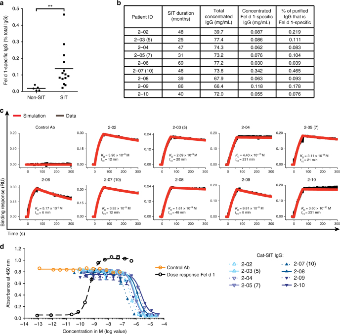
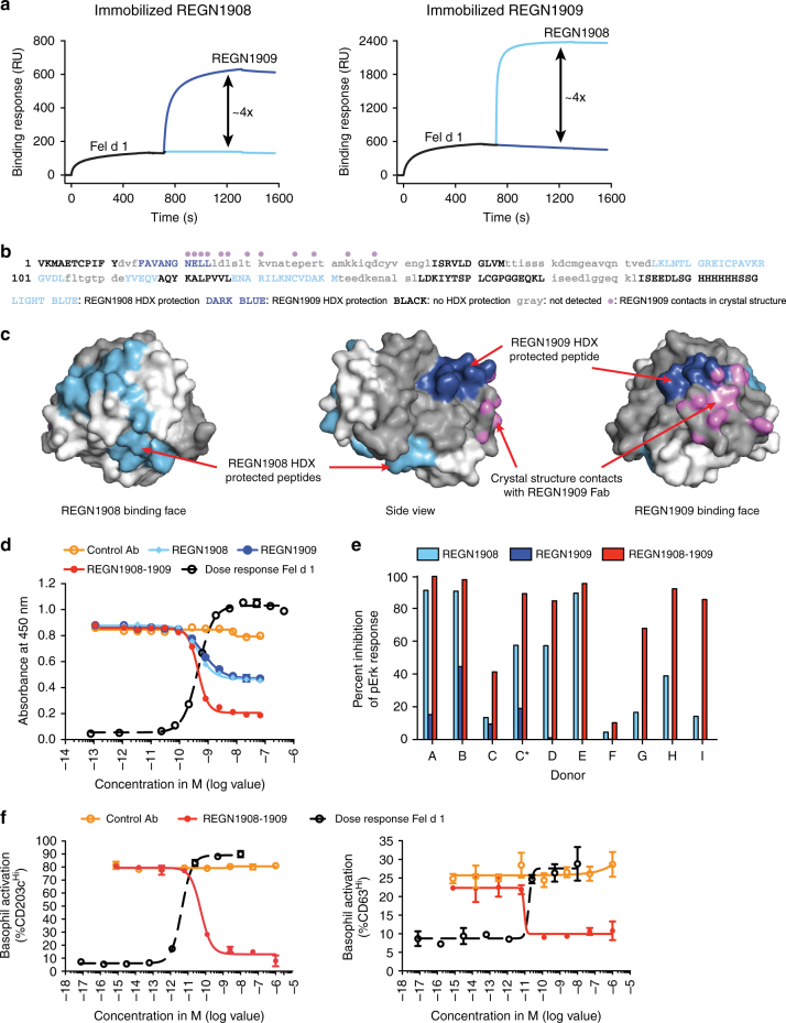
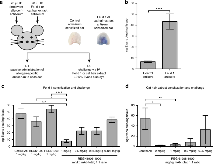
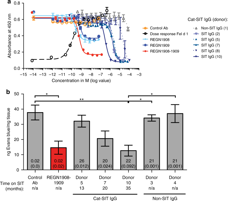
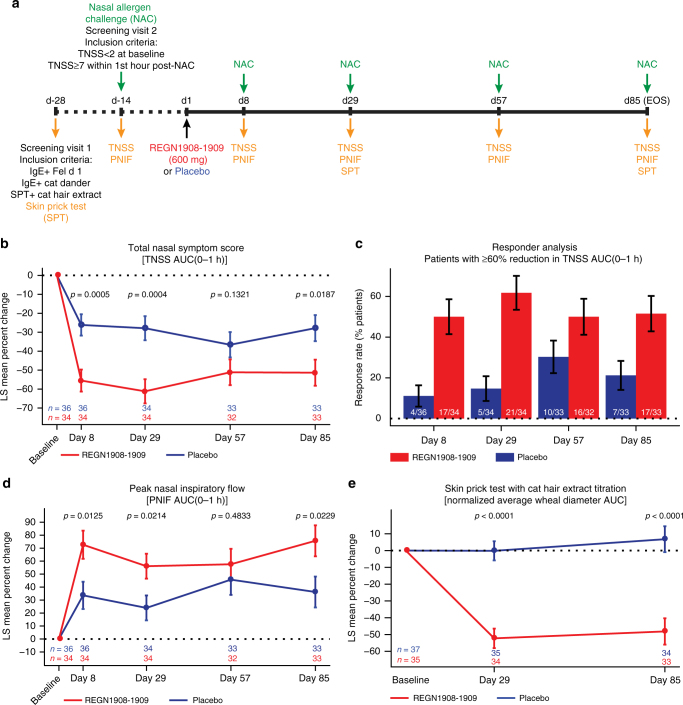
Acute allergic symptoms are caused by allergen-induced crosslinking of allergen-specific immunoglobulin E (IgE) bound to Fc-epsilon receptors on effector cells. Desensitization with allergen-specific immunotherapy (SIT) has been used for over a century, but the dominant protective mechanism remains unclear. One consistent observation is increased allergen-specific IgG, thought to competitively block allergen binding to IgE. Here we show that the blocking potency of the IgG response to Cat-SIT is heterogeneous. Next, using two potent, pre-selected allergen-blocking monoclonal IgG antibodies against the immunodominant cat allergen Fel d 1, we demonstrate that increasing the IgG/IgE ratio reduces the allergic response in mice and in cat-allergic patients: a single dose of blocking IgG reduces clinical symptoms in response to nasal provocation (ANCOVA, p = 0.0003), with a magnitude observed at day 8 similar to that reported with years of conventional SIT. This study suggests that simply augmenting the blocking IgG/IgE ratio may reverse allergy.


Conflict of interest statement

All authors are employees of Regeneron Pharmaceuticals, Inc, and may hold stock and/or stock options in the company.

Figures

Fig. 1
Fig. 1
Specific immunotherapy (SIT) is associated with increased allergen-specific IgGs which have varying affinity between patients and block binding of Fel d 1 to IgE with variable potency. a Fel d 1-specific IgG titers were measured as a percentage of total IgG in cat-allergic Non-SIT (n = 5) and Cat-SIT (n = 14) patient sera by ELISA. Mean (line) and individual patient data representing the average of duplicate wells (dots) are shown. Statistical analysis was assessed by Mann–Whitney two-tailed test. b Individual patient data for samples used in (c) and (d). Patient IDs with parentheses denote three patients who donated blood at two visits. The number in parentheses represents donor ID from the first visit. Binding parameters presented here are based on sample obtained from their second visit. Samples from the first visit were used in the PCA Model, Fig. 4. c Purified and concentrated Cat-SIT-IgG samples from individual patients binding to rFel d 1 was assessed on a HTX biosensor platform. The rFel d 1.mmh was captured on anti-penta-His (HIS1K) Octet biosensors and dipped in samples containing 100 nM of Fel d 1-specific IgG. d Purified and concentrated Cat-SIT IgG from the individual patients shown in b and c were assessed for the ability to block binding of a constant concentration (0.7 nM) of rFel d 1.mmh from binding to plate-captured allergen-specific IgE from one cat-allergic patient in a blocking ELISA. One of two experimental replicates using different IgE donors is shown. The mean and SD from duplicate wells are plotted. Donor 2–06 was not included in the ELISA assay due to volume requirements of the assay. **p< 0.01
Fig. 2
Fig. 2
REGN1908 and REGN1909 simultaneously bind to distinct Fel d 1 epitopes and prevent Fel d 1 binding to Fel d 1-specific IgE. a Non-competitive binding of REGN1908 and REGN1909 was assessed by first injecting nFel d 1 over a CM5 sensor surface immobilized with REGN1908 (left) or REGN1909 (right), followed by the injection of REGN1908 (light blue) or REGN1909 (dark blue). b Using hydrogen/deuterium exchange (HDX), REGN1909 protected a single peptide region in rFel d 1 (dark blue) corresponding to amino acids 32–41 of Chain 2. REGN1908 (light blue) protected two peptide regions corresponding to Chain 1 amino acids 43–47 and 57–71. Light gray lowercase sequence denotes regions not detected by HDX. Violet dots denote residues making contact with REGN1909 Fab as shown in c. c The crystal structure of rFel d 1 bound to the REGN1909 Fab is shown with chain 1 surface colored in white and chain 2 colored in gray. The HDX-protected residues are colored light blue and dark blue as in (b). Atoms making contact (distance <3.5 Å) with the REGN1909 Fab are colored violet. d Representative ELISA using IgE from 1 of 4 donors testing antibody blocking of 0.7 nM rFel d 1.mmh from binding to Fel d 1-specific IgE is shown with mean and SD plotted. e REGN1908, REGN1909, or REGN1908–1909 blocking of 200 pM nFel d 1-induced basophil activation from cat-allergic donors, measured by flow cytometry and shown as percent maximum inhibition of pErk response relative to isotype control antibody. C and C* represent independent blood donations from the same donor 2 months apart. Individual donor and antibody dose–response data are shown in Supplementary Fig. 2. f REGN1908–1909 blocking of 20 pM nFel d 1-induced basophil upregulation of CD203chi (left) or CD63hi (right) was measured by flow cytometry. Average value of duplicate wells and SD are shown. One representative donor out of 8 tested and 1 of 4 are shown for CD203c and CD63 activation, respectively. For combination studies REGN1908–1909 are combined in a 1:1 molar ratio and total antibody is plotted. Flow cytometry gating strategies are shown in Supplementary Fig. 5
Fig. 3
Fig. 3
A combination of REGN1908–1909 inhibits Fel d 1-induced mast cell degranulation in vivo in the passive cutaneous anaphylaxis (PCA) mouse model. a Schematic of the PCA mouse model. b Each mouse (n = 5 in two independent experiments) were sensitized using Fel d 1 antisera and control peanut antisera in the left and right ears, respectively, followed by challenge with 1 μg nFel d 1 one day later. Data from the pooled experiments are expressed as ng of Evans blue extracted per mg of ear tissue from ears sensitized with control antisera or ears sensitized with peanut antisera. Statistical analysis was performed using a two-tailed paired t-test. c Balb/c mice were administered subcutaneous (SC) doses of REGN1908 (n = 15), REGN1909 (n = 15), a combination of REGN1908–1909 (1:1 ratio) at 0.125 mg/kg (n = 15), 0.25 mg/kg (n = 15), 0.5 mg/kg (n = 15), or 1 mg/kg (n = 14), or an IgG4P control antibody (n = 14) 3 days prior to initiating the PCA model using Fel d 1-specific antisera (normalized to 25ng IgE per ear) and 1 µg Fel d 1 challenge. The number per group represents pooled data from three independent experiments. d Balb/c mice were administered SC injections of a combination of REGN1908–1909 (1:1 ratio) at 0.25 mg/kg (n = 5), 0.5 mg/kg (n = 5), or 1 mg/kg (n = 5), or 2 mg/kg (n = 5), or an IgG4P control antibody (n = 5) 3 days prior to initiating the PCA model using cat extract antisera normalized to 25ng IgE per ear and 250 BAU cat hair extract challenge. Results of one experiment are shown. c, d Data are expressed as ng of Evans blue extracted per mg of ear tissue based on the value obtained from each control peanut ear subtracted from the corresponding value from the challenge ear. Statistical significance was determined using the Kruskal–Wallis test followed by the Dunn’s post hoc test (cd). Data are presented as mean ± SE. **** p < 0.0001, ***p < 0.001, **p < 0.01, *p < 0.05
Fig. 4
Fig. 4
REGN1908–1909 blocks Fel d 1 binding to polyclonal Fel d 1-specific IgE more efficiently than natural IgGs from specific immunotherapy (SIT) patients. a In a blocking ELISA, the ability of REGN1908, REGN1909, REGN1908–1909, or Cat-SIT-IgG to block 0.7 nM rFel d 1.mmh from binding to plate-captured Fel d 1-specific IgE from one donor was assessed. A representative experiment using IgE from 1 donor of 3 donors tested is shown. The mean and SD from duplicate wells are plotted. b Balb/c mice were administered subcutaneous (SC) injections of a combination of REGN1908–1909 (1:1 ratio, n = 10), an IgG4P control antibody (n = 10), or concentrated Cat-SIT-IgG from donor 5 (n = 9), donor 7 (n = 10), donor 10 (n = 10) or Non-SIT-IgG from donor 3 (n = 10) or donor 4 (n = 10) prior to the passive cutaneous anaphylaxis model using nFel d 1. Donors 5, 7, and 10 correspond to Donors 2-03(5), 2-05(7), and 2-07(10), respectively in Fig. 1 and Supplementary Table 1. The number per group represents pooled data from two independent experiments. Data are presented as mean ± SE ng of Evans blue extracted per mg of ear tissue is based on the value obtained from each control peanut ear subtracted from the corresponding value from the challenge ear. The total IgG concentration injected (mg), and Fel d 1-specific concentration injected (mg) is represented by the numbers on the bar graphs, with the latter in parentheses. Statistical significance was determined using the Kruskal–Wallis test followed by Dunn’s post hoc test. **p < 0.01 or *p < 0.05
Fig. 5
Fig. 5
REGN1908–1909 blocks the early phase allergic response to cat extract in patients with cat allergy. a Schematic of the study design. b Percent change from baseline in total nasal symptom score (TNSS) AUC(0–1 h); LS mean ± SE is shown for the full analysis set (FAS). c Percentage of patients with ≥60% reduction in TNSS AUC(0–1 h) is shown at each study timepoint. Numbers on the bars indicate the number of patients represented out of the patient number measured at each timepoint. Percentage of patients ± SE is shown for the FAS. d Percent change from baseline in peak nasal inspiratory flow (PNIF) AUC(0–1 h); LS mean ± SE is shown for the FAS. e Percent change from baseline in normalized average wheal diameter AUC is shown from the titrated (100-33,000SQU) cat hair extract skin prick test read 15 min after administration on study days 29 and 85. The skin prick test was not administered on study day 8 or day 57 as per protocol. LS mean ± SE is shown for the safety analysis set. Analyses are based on ANCOVA model with treatment group as a factor and baseline as a covariate. For secondary efficacy and exploratory endpoints no control for multiplicity was performed, therefore p values for all panels are considered nominal

References

    1. Hansen I, Klimek L, Mosges R, Hormann K. Mediators of inflammation in the early and the late phase of allergic rhinitis. Curr. Opin. Allergy Clin. Immunol. 2004;4:159–163. doi: 10.1097/00130832-200406000-00004. - DOI - PubMed
    1. Durham SR, et al. SQ-standardized sublingual grass immunotherapy: confirmation of disease modification 2 years after 3 years of treatment in a randomized trial. J. Allergy Clin. Immunol. 2012;129:717–725.e5. doi: 10.1016/j.jaci.2011.12.973. - DOI - PubMed
    1. Durham SR, et al. Long-term clinical efficacy of grass-pollen immunotherapy. N. Engl. J. Med. 1999;341:468–475. doi: 10.1056/NEJM199908123410702. - DOI - PubMed
    1. Verhoef A, Alexander C, Kay AB, Larche M. T cell epitope immunotherapy induces a CD4+T cell population with regulatory activity. PLoS. Med. 2005;2:e78. doi: 10.1371/journal.pmed.0020078. - DOI - PMC - PubMed
    1. Akdis CA, et al. Epitope-specific T cell tolerance to phospholipase A2 in bee venom immunotherapy and recovery by IL-2 and IL-15 in vitro. J. Clin. Invest. 1996;98:1676–1683. doi: 10.1172/JCI118963. - DOI - PMC - PubMed

Publication types

MeSH terms