. 2018 Apr 12;9(1):1416.
doi: 10.1038/s41467-018-03672-4.

Identification of rare sequence variation underlying heritable pulmonary arterial hypertension

Stefan Gräf  1   2   3 Matthias Haimel  4   5   6 Marta Bleda  4 Charaka Hadinnapola  4 Laura Southgate  7   8 Wei Li  4 Joshua Hodgson  4 Bin Liu  4 Richard M Salmon  4 Mark Southwood  9 Rajiv D Machado  10 Jennifer M Martin  4   5   6 Carmen M Treacy  4   9 Katherine Yates  4   5   6 Louise C Daugherty  5   6 Olga Shamardina  5   6 Deborah Whitehorn  5   6 Simon Holden  11 Micheala Aldred  12 Harm J Bogaard  13 Colin Church  14 Gerry Coghlan  15 Robin Condliffe  16 Paul A Corris  17 Cesare Danesino  18   19 Mélanie Eyries  20 Henning Gall  21 Stefano Ghio  19 Hossein-Ardeschir Ghofrani  21   22 J Simon R Gibbs  23 Barbara Girerd  24 Arjan C Houweling  13 Luke Howard  22 Marc Humbert  24 David G Kiely  16 Gabor Kovacs  25   26 Robert V MacKenzie Ross  27 Shahin Moledina  28 David Montani  24 Michael Newnham  4 Andrea Olschewski  25 Horst Olschewski  25   26 Andrew J Peacock  14 Joanna Pepke-Zaba  9 Inga Prokopenko  22 Christopher J Rhodes  22 Laura Scelsi  19 Werner Seeger  21 Florent Soubrier  20 Dan F Stein  4 Jay Suntharalingam  27 Emilia M Swietlik  4 Mark R Toshner  4 David A van Heel  29 Anton Vonk Noordegraaf  13 Quinten Waisfisz  13 John Wharton  22 Stephen J Wort  22   30 Willem H Ouwehand  5   6 Nicole Soranzo  5   31 Allan Lawrie  32 Paul D Upton  4 Martin R Wilkins  22 Richard C Trembath  8 Nicholas W Morrell  33   34
Affiliations

Identification of rare sequence variation underlying heritable pulmonary arterial hypertension

Stefan Gräf et al. Nat Commun. .

Abstract

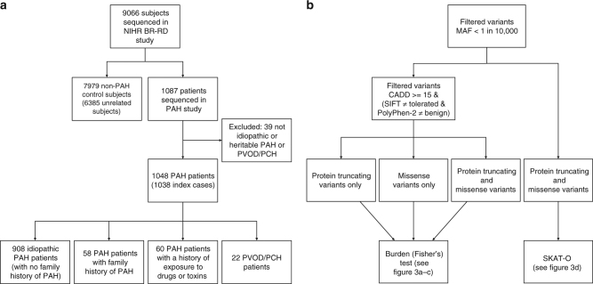
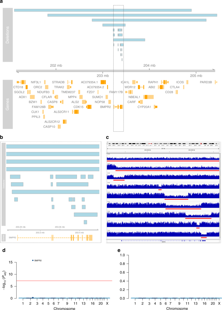
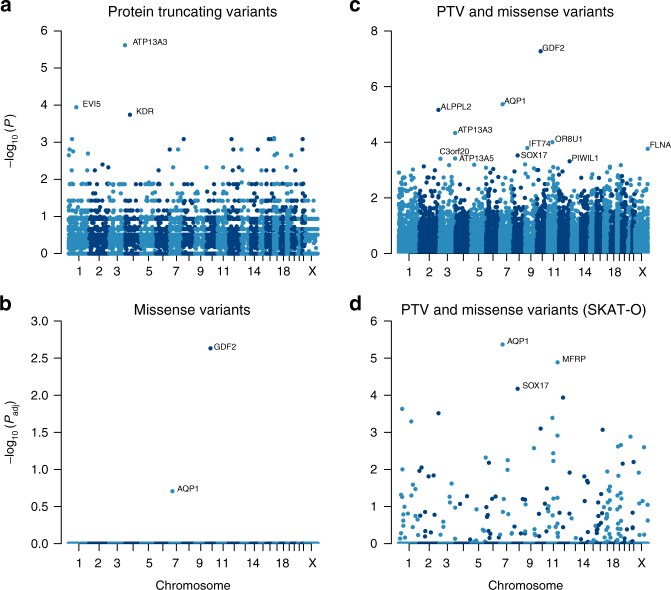
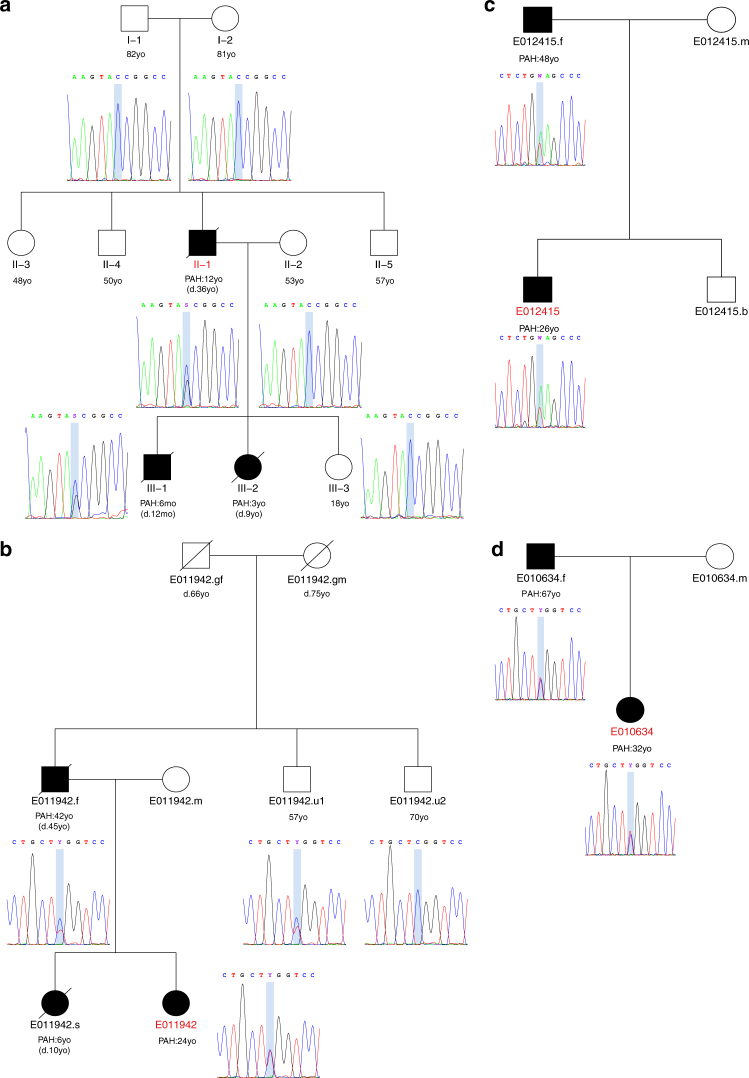
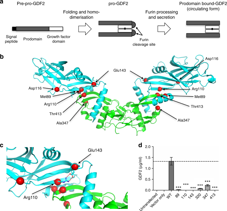
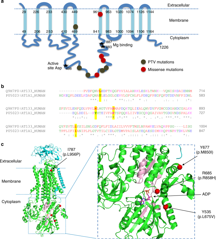
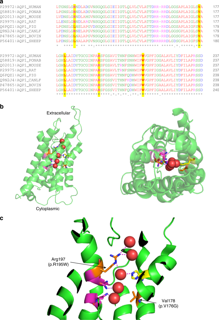
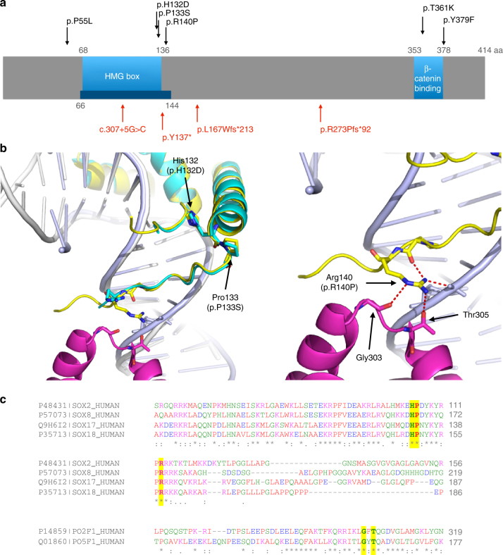
Pulmonary arterial hypertension (PAH) is a rare disorder with a poor prognosis. Deleterious variation within components of the transforming growth factor-β pathway, particularly the bone morphogenetic protein type 2 receptor (BMPR2), underlies most heritable forms of PAH. To identify the missing heritability we perform whole-genome sequencing in 1038 PAH index cases and 6385 PAH-negative control subjects. Case-control analyses reveal significant overrepresentation of rare variants in ATP13A3, AQP1 and SOX17, and provide independent validation of a critical role for GDF2 in PAH. We demonstrate familial segregation of mutations in SOX17 and AQP1 with PAH. Mutations in GDF2, encoding a BMPR2 ligand, lead to reduced secretion from transfected cells. In addition, we identify pathogenic mutations in the majority of previously reported PAH genes, and provide evidence for further putative genes. Taken together these findings contribute new insights into the molecular basis of PAH and indicate unexplored pathways for therapeutic intervention.


Conflict of interest statement

The authors declare no competing interests.

Figures

Fig. 1
Fig. 1
Flow diagrams illustrating a the composition of the NIHR BioResource—Rare Diseases (NIHR BR-RD) PAH study and b the analysis strategy to identify novel PAH disease genes. a The study comprised 1048 adult cases (aged 16 or over) attending specialist pulmonary hypertension centres from the UK (n = 731), and additional cases from France (n = 142), The Netherlands (n = 45), Germany (n = 82) and Italy (n = 48). b A series of case-control comparisons including and excluding cases with variants in previously reported disease genes were undertaken using complementary filtering strategies
Fig. 2
Fig. 2
Analysis of copy number deletions. a Deletions affecting the BMPR2 locus in 23 PAH cases. Genes are indicated in orange and labelled with their respective gene symbol. Deletions are drawn as blue boxes above the genome axis (grey) showing the genomic position on chromosome 2. The grey box highlights the location of BMPR2. b Locus zoom on BMPR2 highlighting the focal deletions affecting one or more exons. c WGS coverage profiles of a selected set of smaller and larger deletions, visualised with the Integrative Genomics Viewer (IGV), with deletions highlighted by red bars. d and e Manhattan plots of the genome-wide case-control comparison of large deletions. In d, all subject are considered. In e, subject with larger deletions affecting the BMPR2 locus are excluded. The adjusted P value threshold of 5 × 10−8 for genome-wide significance is indicated by the red line
Fig. 3
Fig. 3
Manhattan plots of the rare variant analyses, having excluded cases carrying rare variants in previously established PAH genes. Filtered variants were grouped per gene. We tested for an excess of variants in PAH cases within genes using Fisher’s exact test. The negative decadic logarithm of unadjusted or adjusted P-values are plotted against the chromosomal location of each gene. a Burden test of rare PTVs. b Burden test of rare deleterious missense variants. c Burden test combining rare PTVs and likely deleterious missense variants. d SKAT-O test of rare PTVs and missense variants
Fig. 4
Fig. 4
Pedigree structures and analysis of familial transmission of variants in AQP1 and SOX17. a Individual II.1 harbours a heterozygous de novo SOX17 c.411 C > G (p.Y137*) PTV resulting in a premature termination codon, which has been transmitted to the affected male (III.1). No unaffected family members carry the variant. No sample was available from subject III.2. b Proband E011942 has inherited a heterozygous AQP1 c.583 C > T (p.R195W) missense variant from her affected father. No sample was available from the affected sister of the proband. The younger healthy uncle of the index case also carries the AQP1 variant. No samples or further clinical information was available for the grandparents, who were not known to have cardiopulmonary disease. c Both the proband E012415 and her father are affected and carry the rare AQP1 c.527 T > A (p.V176E) missense variant. There was no further information available about the siblings of the father. d Subject E010634 has inherited the heterozygous AQP1 c.583 C > T (p.R195W) missense variant from her affected father. No rare variants in previously reported PAH genes were identified in any of theses families. Index cases are highlighted in red. d death, mo months old, yo years old
Fig. 5
Fig. 5
Structural analysis of GDF2 mutations. a Schematic diagram of GDF2 processing. The pre-pro-protein is processed into the mature growth factor domain (GFD) bound to the prodomain upon secretion. b Plot of GDF2 mutations found only in PAH cases superimposed on the structure of prodomain bound GDF2 (PDB: 4YCG). The GDF2 growth factor domain is shown in green and the prodomain in cyan. c Magnified view of the Arg110 and Glu143 mutations. The wild-type amino acids make double salt bridges to stabilise the prodomain conformation at the interface between the growth factor domain and prodomain. The E143K and R110W mutations both disrupt these interactions, destabilising the interaction between the growth factor domain and prodomain. d GDF2 levels secreted into supernatants of HEK293T cells transfected with likely pathogenic variants found in PAH cases, compared with wild-type GDF2 and cells transfected with an empty vector. ***P < 0.001 by ANOVA
Fig. 6
Fig. 6
Structural analysis of ATP13A3 mutations. a Topology of ATP13A3, plotted according to UniProtKB Q9H7F0. Frameshift and stop-gained mutations identified in PAH cases are shown as khaki circles, and missense mutations as red circles. Frameshift/stop-gained mutations are predicted to truncate the protein prior to the catalytic domain and essential Mg binding sites, leading to loss of ATPase activity. b Sequence alignment of ATP13A3 with ATP1A1 (P05024), of which the high resolution structure was used for the structural analysis in c. The conserved regions of ATP13A3 and ATP1A1, essential for ATPase activity, show good alignment (data not shown). Only regions containing the missense PAH mutations are shown, with positions of the four missense mutations highlighted in yellow above the sequences. c Structural analysis of the 4 PAH missense mutations plotted on the ATP1A1 crystal structure based on the sequence alignment in b (PDB: 3wgu). Green: α subunit (P05024), cyan: β subunit (P05027), grey: γ-subunit transcript variant a (Q58k79). Y535, Y677, R685 and I787 are the numbering in ATP1A1. Positions of the four missense mutations found in PAH are labelled and highlighted by red circles. d Magnified view of the cytoplasmic region of the ATPase, showing the presence of ADP at the active site. The conserved regions essential for ATPase activity are shown in light pink. The L675V and R858H mutations are located close to the ATP catalytic region
Fig. 7
Fig. 7
Structural analysis of AQP1 mutations. a Multiple sequence alignment of human AQP1 with seven other mammals. The bovine AQP1 has the high resolution (2.2 Å) published structure. Mutations identified in PAH cases are highly conserved and highlighted in yellow. b Crystal structure of bovine AQP1 (PDB: 1j4n). Left: side view; right: top view from the extracellular direction. AQP1 is shown as a semi-transparent cartoon and five water molecules in the water channel are shown as red spheres. Key residues lining the water channels are represented with stick structures. c Magnified view of the water channel, with H-bonds connected to water molecules in the channel highlighted. Two asparagine-proline-alanine (NPA) motifs, essential for the water transporting function of AQP1, are shown in magenta. Conserved His180 that constricts the water channel is shown in yellow. Mutations found in PAH cases, Arg195Trp and Val176Glu, are labelled and shown as orange stick structures. Arg195 and His180 are highly conserved in the known water channels and are strong indicators of water channel specificity. Arg195Trp and Val176Glu mutations are predicted to disrupt the conformation of this conserved water channel
Fig. 8
Fig. 8
Structural analysis of SOX17 mutations. a Schematic diagram of human SOX17 (Q9H6I2), based on UniProtKB annotation, and published reports. Red arrows indicate PTVs and black arrows indicate missense mutations identified in PAH patients. The blue bar illustrates the region that is covered in the crystal structure (PDB: 3F27). The ability of SOX17 to activate transcription of target genes correlates with binding to β-catenin. As illustrated, all PTVs lead to a loss of the β-catenin binding region. Two missense mutations are located within and very close to the minimum β-catenin binding regions, and both are highly conserved, indicating they are likely to be important for β-catenin binding. b Structural analysis of HMG domain missense mutations found in PAH patients. Left, Superposition of SOX17/DNA structure (Sox17: cyan, DNA: grey) onto SOX2/DNA/Oct1 structure (PDB: 1GT0, Sox2: yellow, Oct1: magenta, DNA: light blue). Right: Magnified view of the interactions around Arg140 in the SOX2/DNA/Oct structure. Arg140 in SOX2 makes multiple H-bond interactions and mutating this Arg in SOX2 abolishes the interaction with transcription factors Pax6 and Oct4. SOX2 and SOX17 both bind to Oct4 and SOX17 K122E mutant can replace SOX2 in maintaining stem cell pluripotency, indicating this region in SOX17 may interact with Oct4, similar to SOX2. The three missense mutations in SOX17 will likely disrupt interaction with Oct4. c Supporting the analysis in b, sequence alignment shows that the HMG domain of SOX2 (P48431) and SOX17 as well as SOX8 (P57073) and SOX18 (P35713) share high sequence identity and the three mutations found in PAH (highlighted in yellow) are highly conserved emphasising their functional importance. Similarly, the Gly and Thr that interact with Arg140 in SOX2 (highlighted in yellow) are also conserved between Oct1 (PO2F1) and Oct4 (PO5F1)
Fig. 9
Fig. 9
Immunolocalisation of AQP1, ATP13A3 and SOX17 in normal and PAH lung. The typical histological findings (haematoxylin and eosin staining) of concentric vascular lesions with associated plexiform lesions are shown (a). Higher magnification images of plexiform lesion (b), with frequent endothelialised channels (c; anti-CD31) surrounded by myofibroblasts (d; anti-SMα). Additional high magnification images demonstrating endothelial expression of ATP13A3 (e), AQP1 (f) and SOX17 (g) in PAH lung. Controls lung sections demonstrating predominantly endothelial expression of ATP13A3 (h), AQP1 (i) and SOX17 (j). (Scale bars = 50 µm)
Fig. 10
Fig. 10
Functional studies of novel genes. ac Expression of a AQP1, b ATP13A3 and c SOX17 mRNA in human pulmonary artery smooth muscle cells, pulmonary artery endothelial cells and blood outgrowth endothelial cells (BOECs) (n = 4 biological replicates of each). Relative expression of each transcript was normalised to three reference genes, ACTB, B2M and HPRT. d Proliferation of BOECs in 5% FBS over 6 days. Cells were transfected with DharmaFECT1 alone (DH1), siATP13A3 or non-targeting siRNA control (siCP) e, f Quantification of apoptosis in BOECs, defined as Annexin V+/PI− cells, in BOECs transfected with siATP13A3 or siCP in complex with DH1 followed by 24 h treatment with 0.1% FBS or 5% FBS (n = 4 biological repeats). f Measurement of apoptosis via Caspase-Glo 3/7 activity measurements in BOECs transfected with siATP13A3 or siCP in complex with DH1, followed by 16 h treatment in 0.1% FBS or 5% FBS. Data are from a single experiment (n = 4 wells) representative of 3 biological repeats. Data were analysed using a One-way analysis of variance with post hoc Tukey’s test for multiple comparisons in d and f. Data were analysed using a repeated measures One-way analysis of variance with post hoc Tukey’s for multiple comparisons in e. *P < 0.05, **P < 0.01 within treatment groups. ###P < 0.001 for effect of ligand against control for same transfection condition

References

    1. Wagenvoort C. A. The pathology of primary pulmonary hypertension. J. Pathol.101, Pi (1970) - PubMed
    1. McGoon MD, et al. Pulmonary arterial hypertension: epidemiology and registries. J. Am. Coll. Cardiol. 2013;62(25 Suppl):D51–D59. doi: 10.1016/j.jacc.2013.10.023. - DOI - PubMed
    1. McLaughlin VV, Shillington A, Rich S. Survival in primary pulmonary hypertension: the impact of epoprostenol therapy. Circulation. 2002;106:1477–1482. doi: 10.1161/01.CIR.0000029100.82385.58. - DOI - PubMed
    1. Lane KB, et al. Heterozygous germline mutations in BMPR2, encoding a TGF-beta receptor, cause familial primary pulmonary hypertension. Nat. Genet. 2000;26:81–84. doi: 10.1038/79226. - DOI - PubMed
    1. Deng Z, et al. Familial primary pulmonary hypertension (gene PPH1) is caused by mutations in the bone morphogenetic protein receptor-II gene. Am. J. Hum. Genet. 2000;67:737–744. doi: 10.1086/303059. - DOI - PMC - PubMed

Publication types

MeSH terms