. 2018 Apr 12;9(1):1420.
doi: 10.1038/s41467-018-03704-z.

Epigenetic control of IL-23 expression in keratinocytes is important for chronic skin inflammation

Affiliations

Epigenetic control of IL-23 expression in keratinocytes is important for chronic skin inflammation

Hui Li et al. Nat Commun. .

Abstract

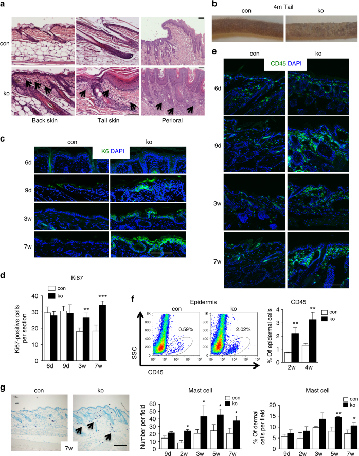
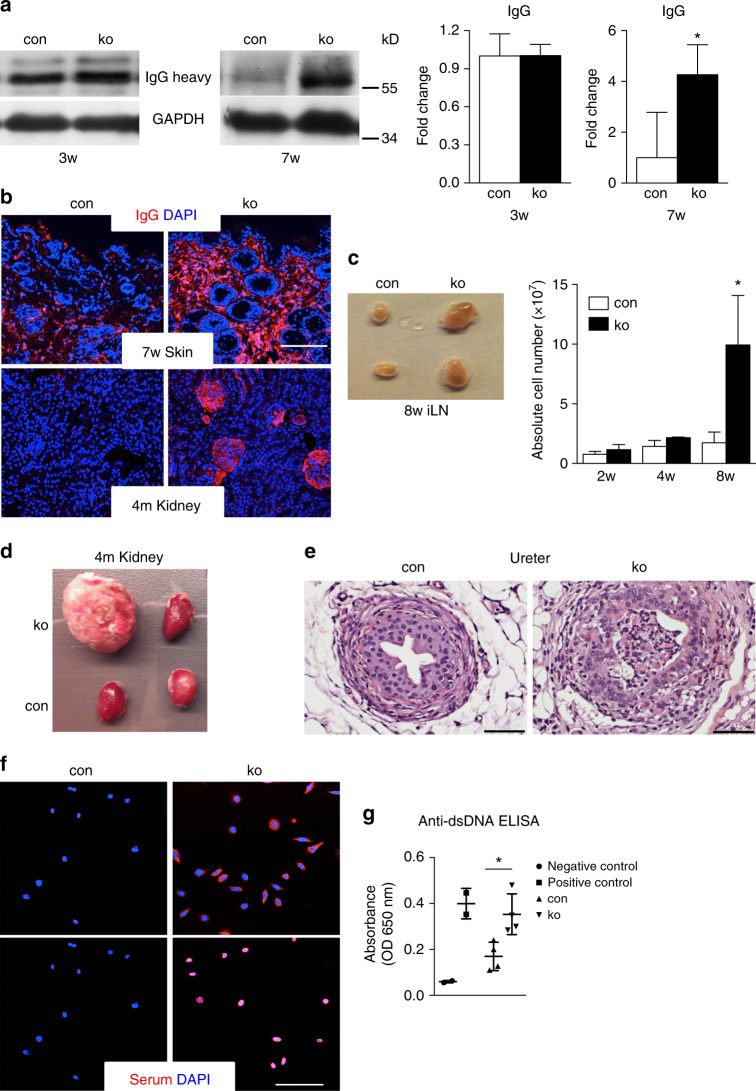
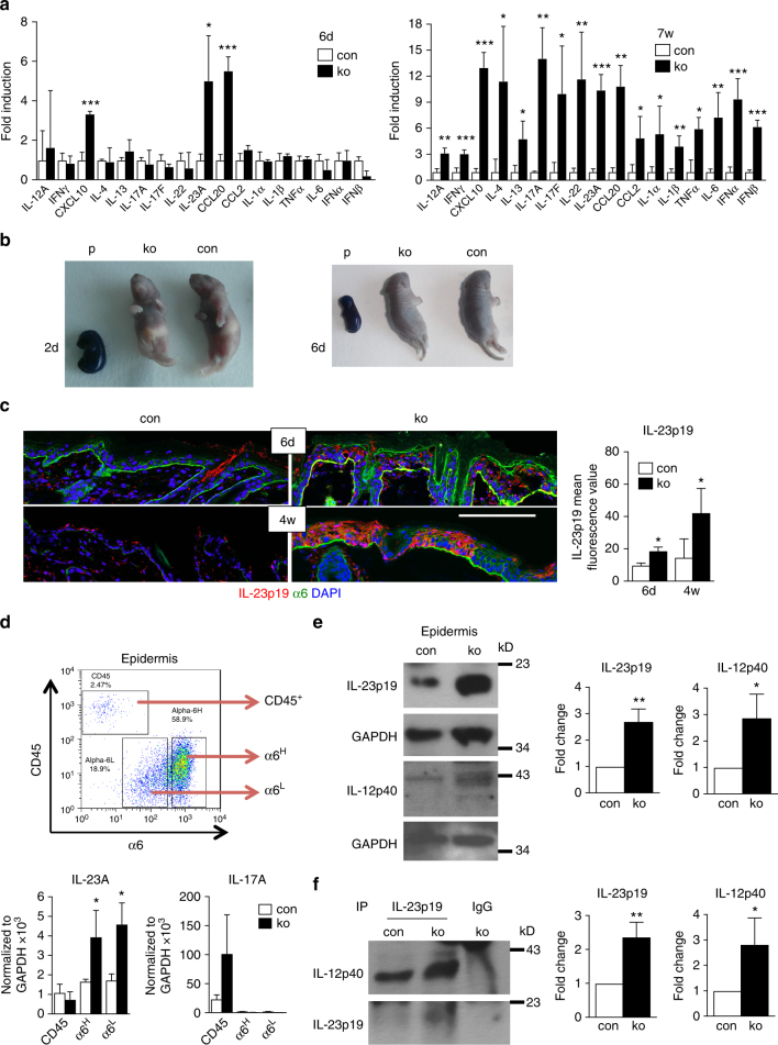
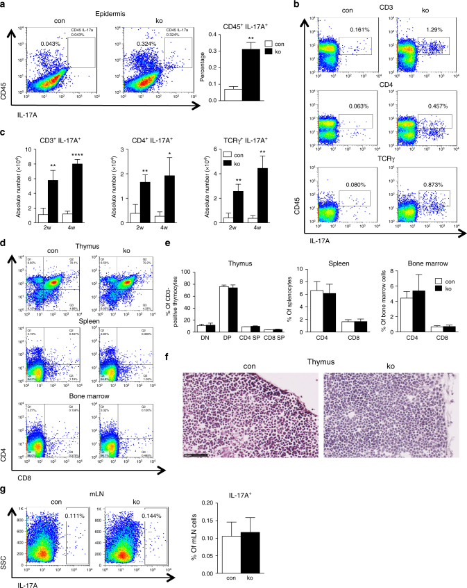
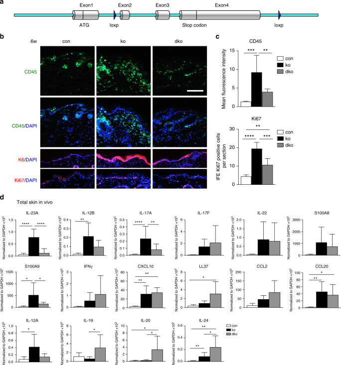
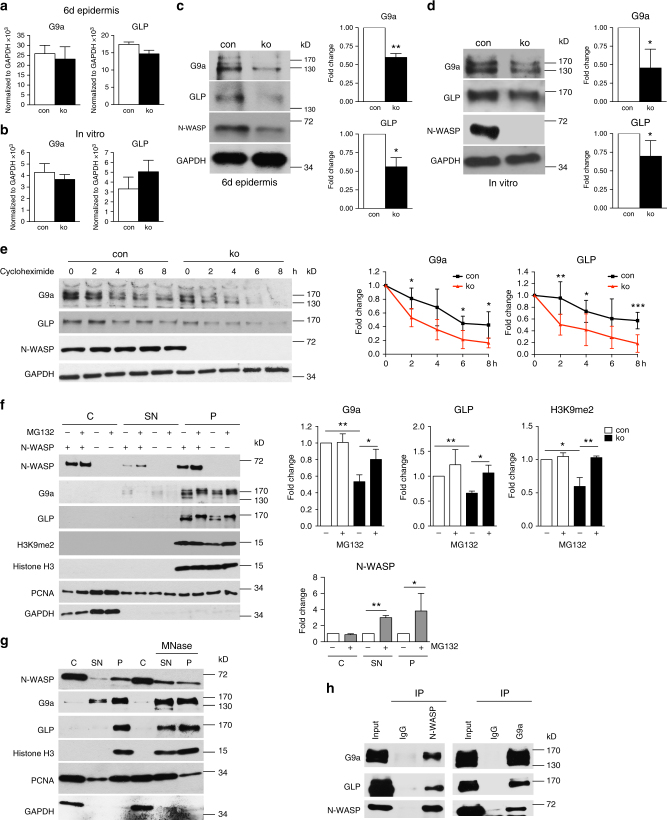
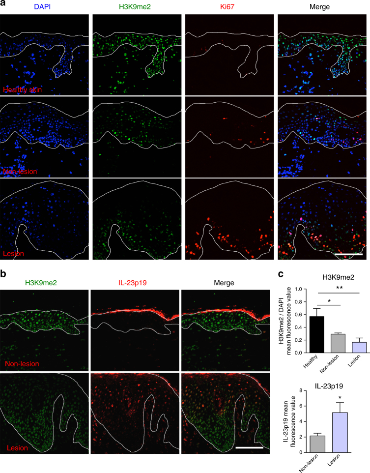
The chronic skin inflammation psoriasis is crucially dependent on the IL-23/IL-17 cytokine axis. Although IL-23 is expressed by psoriatic keratinocytes and immune cells, only the immune cell-derived IL-23 is believed to be disease relevant. Here we use a genetic mouse model to show that keratinocyte-produced IL-23 is sufficient to cause a chronic skin inflammation with an IL-17 profile. Furthermore, we reveal a cell-autonomous nuclear function for the actin polymerizing molecule N-WASP, which controls IL-23 expression in keratinocytes by regulating the degradation of the histone methyltransferases G9a and GLP, and H3K9 dimethylation of the IL-23 promoter. This mechanism mediates the induction of IL-23 by TNF, a known inducer of IL-23 in psoriasis. Finally, in keratinocytes of psoriatic lesions a decrease in H3K9 dimethylation correlates with increased IL-23 expression, suggesting relevance for disease. Taken together, our data describe a molecular pathway where epigenetic regulation of keratinocytes can contribute to chronic skin inflammation.


Conflict of interest statement

The authors declare no competing interests.

Figures

Fig. 1
Fig. 1
Chronic skin inflammation in mice with a keratinocyte-restricted deletion of N-WASP. a H&E-stained back, tail, and perioral skin sections of 7-week-old mice (n: 6/6). Arrows indicate increased dermal cellularity. b Representative image of tails of 4-month (4m)-old control and ko mice. c K6 immunofluorescence of back skin section of at indicated ages (n: 4/4, mean ± SD, two-tailed unpaired t-test). d Quantification of Ki67+ cells in back skin epidermis sections (n: 4/4, mean ± SD, two-tailed unpaired t-test). e CD45 immunofluorescence of back skin at the indicated ages (n: 4/4). f FACS for CD45+ cells in back skin epidermis and quantification (n: 3/3, mean ± SD, two-tailed unpaired t-test). g Representative toluidine blue staining for mast cells and quantification of mast cell infiltration in numbers per field and ratio of dermal cells per field (n: 3/3, two-tailed unpaired t-test). Arrows indicate mast cells. Scale bars (a, c, e, g): 100 µm (*p ≤ 0.05; **p ≤ 0.01; ***p ≤ 0.001)
Fig. 2
Fig. 2
N-WASP knockout keratinocytes show multi-organ inflammation and autoimmunity. a Immunoblot analysis of IgG deposition in back skin at indicated ages. All normalized to GAPDH (n > = 3, mean ± SD, two-tailed unpaired t-test). b Representative immunofluorescence of IgG deposition of back skin and kidney at indicated ages (n: 3/3, mean ± SD, two-tailed unpaired t-test). Scale bar: 100 µm. c Representative image of inguinal lymph nodes (iLN) of con and ko mouse and quantification of cellularity of iLN at indicated ages (n: 3/3, mean ± SD, two-tailed unpaired t-test). d Representative image of kidneys of 4-month (4m)-old control and ko mice. e Representative of H&E staining of ureter. Scale bar: 50 µm. f Representative immunofluorescence staining of serum from con and ko (n: 8/8). g Detection of anti-dsDNA antibodies in serum (1:200) by ELISA (n: 4/4; mean ± SD, two-tailed unpaired t-test; *p ≤ 0.05)
Fig. 3
Fig. 3
Increased cytokine expression in N-WASP-null skin. a qRT-PCR analysis of indicated genes from back skin RNA of con and ko at indicated ages (n > = 3/3, two-tailed unpaired t-test). All normalized to GAPDH. b Representative toluidine blue staining for barrier defect in con and ko mice at indicated ages (P: E15.5 embryos used as positive controls; 2 days (2d) n: 6/5, 6 days(6d) n: 4/5, 3 independent experiments). c Representative immunofluorescence staining and quantification of IL-23p19 of con and ko back skin at indicated ages (n: 3/3, two-tailed paired t-test). Scale bar: 100 µm. d Representative sorting strategy of CD45+, α6H, and α6L cells from con and ko epidermal cells and qRT-PCR analysis of IL-23A and IL-17A expression in sorted cells (n: 3/3, mean ± SD, two-tailed unpaired t-test, 3 independent sortings). e Representative immunoblots and quantification of indicated proteins from epidermal lysates of 7–8-week old con and ko mice (n: 4/4, mean ± SD, two-tailed unpaired t-test). f Representative immunoblots and quantification of indicated proteins of co-precipitations of IL-23p19 with IL-12p40 from epidermal lysates of 7–8-week-old con and ko mice (n: 4/4, mean ± SD, two-tailed unpaired t-test; *p ≤ 0.05; **p ≤ 0.01; ***p ≤ 0.001)
Fig. 4
Fig. 4
Increased IL-17A-producing cells in N-WASP ko mice. a Representative and quantification of intracellular FACS analysis of IL-17A+ cells in epidermis (n: 3/3, mean ± SD, two-tailed unpaired t-test). b, c Representative and quantification of intracellular FACS analysis of IL-17A+ cells in iLN (n: 3/3, mean ± SD, two-tailed unpaired t-test). d, e Representative and quantification of CD4, CD8 distribution in thymus, spleen, and bone marrow from 9-day-old con and ko mice (n: 3/3, two-tailed unpaired t-test). f Representative H&E staining of thymus from 9-day-old con and ko mice (n: 3/3). Scale bar: 50 µm. g Representative and quantification of intracellular FACS analysis of IL-17A+ cells in mLN (n: 6/6, mean ± SD, two-tailed unpaired t-test; *p ≤ = 0.05; **p ≤ = 0.01; ****p ≤ = 0.0001)
Fig. 5
Fig. 5
Keratinocyte-derived IL-23p19 is important for skin inflammation in N-WASP knockout. a Strategy of loxP insertion in mouse IL-23A gene. b, c CD45, K6, Ki67 immunofluorescence of back skin section from 6-week-old con, ko, and dko (b), quantification of mean fluorescence intensity of CD45, and quantification of Ki67-positive cells in interfollicular epidermis (IFE) (n: 5/5/5, mean ± SD, one-way ANOVA, with Tukey’s multiple comparisons). Scale bar: 100 µm. d qRT-PCR analysis of indicated genes from back skin RNA 6–8-week-old of con, ko, and dko mice. All normalized to GAPDH (n > = 6/6/6, mean ± SD, one-way ANOVA, with Tukey’s multiple comparisons; *p ≤ 0.05; **p ≤ 0.01; ***p ≤ 0.001; ****p ≤ 0.0001)
Fig. 6
Fig. 6
IL-23A expression is increased in N-WASP-null keratinocytes. a qRT-PCR analysis of indicated genes in FACS purified primary keratinocytes from 6-week-old mice (n: 4/4/4, mean ± SD, two-tailed unpaired t-test, 2 independent sorting). b qRT-PCR analysis of indicated cytokine mRNA expression in primary keratinocytes. All normalized to GAPDH (n: 3/3, mean ± SD, two-tailed unpaired t-test). c Primary keratinocytes were differentiated with 2 mM CaCl2 (High Ca2+) for 48 h and then analyzed by qRT-PCR for IL-23A expression, compared to untreated cells (Low Ca2+; n: 3/3, mean ± SD, two-tailed unpaired t-test). d qRT-PCR analysis of IL-23A mRNA expression in primary keratinocytes in response to 24 h treatment with indicated inhibitors. All normalized to GAPDH (n: 3/3, mean ± SD, two-tailed unpaired t-test). e Representative immunofluorescence of phalloidin in con and ko back skin. Scale bar: 100 µm. f Quantification of FACS mean fluorescence intensity of phalloidin in con and ko primary keratinocyte (n: 3/3, mean ± SD, two-tailed unpaired t-test). g qRT-PCR analysis of IL-23A mRNA expression in primary keratinocytes treated with indicated inhibitors for 24 h. All normalized to GAPDH. The right panel shows the same data, but normalized to the respective DMSO-treated samples, thus showing the relative effect of the treatment as fold change (n: 3/3, mean ± SD, two-tailed unpaired t-test; *p ≤ 0.05; **p ≤ 0.01; ***p ≤ 0.001; ****p ≤ 0.0001)
Fig. 7
Fig. 7
N-WASP represses IL-23p19 expression in keratinocytes by regulating H3K9 dimethylation. a, b Immunoblot analysis of H3K9me2 level in con and ko primary keratinocytes treated with BIX01294 (a) or IOX-1 (b) for 24 h (n: 3/3, mean ± SD, one-way ANOVA, with Tukey’s multiple comparisons). c Immunoblot for H3K9me2 in epidermis of 6-day-old mice (n: 3/3, mean ± SD, two-tailed unpaired t-test). d H3K9me2 IF of back skin of 6-day-old mice (n: 3/3). Scale bar: 100 µm. e qRT-PCR analysis of IL-23A mRNA expression in sorted interfollicular epidermal keratinocytes from con and G9a knockout. All normalized to GAPDH (n: 4/4, mean ± SD, two-tailed unpaired t-test). f ChIP of primary keratinocytes for H3K9me2 with qPCR for indicated regions of IL-23 promoter (n: 7/7, mean ± SD, two-tailed unpaired t-test). g, h Primary N-WASP fl/fl cells lentivirally transduced with GFP or Cre-GFP were analyzed by western blot for indicated genes (g) and by qRT-PCR for IL-23A expression (h; n: 4/4, mean ± SD, two-tailed unpaired t-test; *p ≤0.05; **p ≤ 0.01; ***p ≤ 0.001)
Fig. 8
Fig. 8
N-WASP co-precipitates with and regulates the stability of the H3K9 methyltransferases G9a and GLP. Expression of G9a and GLP mRNA (a, b) and protein (c, d) in epidermis of 6-day-old mice (a, c) and in primary keratinocytes (b, d). (n (a, b, c): 3/3; n (d): 5/5). e immunoblot analysis of protein stability of G9a and GLP in primary keratinocytes in the presence of cycloheximide (G9a n: 4/4, GLP n: 6/6, mean ± SD, two-tailed unpaired t-test). f Cell fractionation of keratinocytes after 90 min of treatment with the proteasome inhibitor MG-132 followed by immunoblot for indicated proteins, the indicated protein levels were quantified on the right (n: 3/3, mean ± SD, two-tailed unpaired t-test). g Cell fractionation of primary keratinocytes with treatment of the pellet fraction with micrococcal nuclease followed by immunoblot for indicated proteins (MNase) (n: 3). h Co-precipitation of N-WASP with G9a and GLP in primary keratinocyte nuclear lysates (n: 3). C: cytosolic fraction, SN: soluble nuclear fraction, P: pallet (*p ≤ 0.05; **p ≤ 0.01; ***p ≤ 0.001)
Fig. 9
Fig. 9
TNF regulates IL-23A expression by phosphorylating N-WASP and degradation of G9a/GLP complex. a qRT-PCR analysis of IL-23A mRNA expression in con and ko primary keratinocytes treated for 4 h with TNFα (n: 3/3, mean ± SD, two-tailed unpaired t-test). b FACS analysis of surface TNF-RI in primary keratinocytes (n: 3/3, two-tailed unpaired t-test). c Primary keratinocytes were treated for indicated times with TNFα and then tested by immunoblot for expression of indicated proteins (n: 4/4). d Primary keratinocytes were treated for indicated time with TNF, fractionated, and analyzed by immunoblot for indicated proteins (C: cytosolic fraction, S300: soluble nuclear fraction extracted by 300 mM NaCl, P300: pellet after 300 mM NaCl extraction, SE: short exposure, LE: long exposure; n: 3). e ChIP of primary keratinocytes for H3K9me2 with qRT-PCR for indicated regions of IL-23A promoter after treatment with TNFα for indicated times (n: 5/5, mean ± SD, two-tailed unpaired t-test). f qRT-PCR analysis of IL-23A mRNA expression in con and ko primary keratinocytes pretreated with or without IOX-1 for 24 h and then stimulated with or without TNFα for 4 h as indicated. All normalized to GAPDH (n: 4/4, mean ± SD, two-tailed unpaired t-test; *p ≤ 0.05; **p ≤ 0.01; ***p ≤ 0.001)
Fig. 10
Fig. 10
Decreased H3K9me2 in psoriatic lesions correlating with increased IL-23p19 expression. a, b H3K9me2 staining (a) and quantification (b) in healthy skin, non-lesioned and lesioned psoriatic skin (n: 3/3, mean ± SD, one-way ANOVA, with Tukey’s multiple comparisons). c H3K9me2 and IL-23p19 staining in non-lesioned and lesioned psoriatic skin (n: 3/3). Scale bars: 100 µm (a, c). (*p ≤ = 0.05; **p ≤ = 0.01)

Similar articles

Cited by

References

    1. Nestle FO, Kaplan DH, Barker J. Psoriasis. N. Engl. J. Med. 2009;361:496–509. doi: 10.1056/NEJMra0804595. - DOI - PubMed
    1. Kim J, Krueger JG. The immunopathogenesis of psoriasis. Dermatol. Clin. 2015;33:13–23. doi: 10.1016/j.det.2014.09.002. - DOI - PubMed
    1. Campa M, Menter A. A review of emerging IL-17 inhibitors in the treatment of psoriasis focusing on preclinical through phase II studies. Expert Opin. Investig. Drugs. 2016;25:1337–1344. doi: 10.1080/13543784.2016.1237502. - DOI - PubMed
    1. Suarez-Farinas M, et al. Alopecia areata profiling shows TH1, TH2, and IL-23 cytokine activation without parallel TH17/TH22 skewing. J. Allergy Clin. Immunol. 2015;136:1277–1287. doi: 10.1016/j.jaci.2015.06.032. - DOI - PubMed
    1. Wong CK, et al. Hyperproduction of IL-23 and IL-17 in patients with systemic lupus erythematosus: implications for Th17-mediated inflammation in auto-immunity. Clin. Immunol. 2008;127:385–393. doi: 10.1016/j.clim.2008.01.019. - DOI - PubMed

Publication types

MeSH terms