. 2018 Apr 12;8(1):5863.
doi: 10.1038/s41598-018-21807-x.

Hydrogen sulfide inhibits epithelial-mesenchymal transition in peritoneal mesothelial cells

Affiliations

Hydrogen sulfide inhibits epithelial-mesenchymal transition in peritoneal mesothelial cells

Shengnan Cheng et al. Sci Rep. .

Abstract

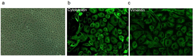
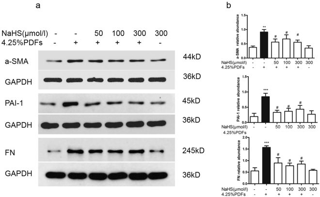
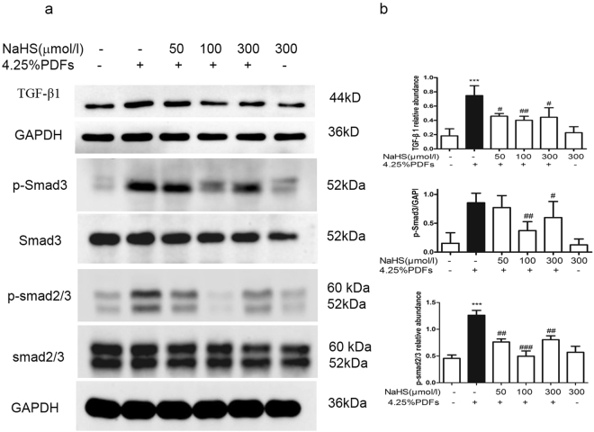
Peritoneal fibrosis (PS) determines the long-term outcome of peritoneal dialysis (PD). We previous confirmed that hydrogen sulfide (H2S) inhibited PS, but its cellular mechanism was not fully elucidated. Epithelial-mesenchymal transition (EMT) of mesothelial cells (MCs) is an important cellular event of PS, we therefore investigated whether EMT can be affected by H2S in MCs. Rats were treated with 4.25% -glucose PD fluids plus lipopolysaccharide for 28 days to produce PS, and NaHS (56 μg/kg.d) was given simultaneously. NaHS (56 μg/kg.d) reduced the deposition of collagen in the submesothelial zone compared with the PS group. In primarily cultured rat MCs, 4.25% -glucose PD fluid induced EMT in MCs featured as loss of ZO-1 and Cytokeratin, and increase of α-SMA, plasminogen activator inhibitor 1, fibronectin and TGF-β1 proteins. PD fluid also increased IL-6 and monocyte chemotactic protein-1 mRNA expressions as well as the phosphorylation of Smad2/3 and Smad3. NaHS (50-300 μmol/L) reversed the above alterations with the optimal dose at 100 μmol/L. Thus, exogenous H2S improves PS by inhibiting EMT in MCs. The anti-EMT effect of H2S is associated with the inhibition of inflammation and TGF-β1-Smad signal pathway.


Conflict of interest statement

The authors declare no competing interests.

Figures

Figure 1
Figure 1
Masson-trichrome staining of rat peritoneal tissue. (a) On the 28th day, the fibrotic thickness of the rat peritoneum in the PD group was significantly increased compared with the control group. Administration of NaHS (56 µg/kg/day) in the PD rats markedly reduced the deposition of collagen fibers. The thickness of collagen fibers (blue) of the NaHS group and the control group was comparable. Magnification × 200. (b) Quantitative analysis of the collagen thickness (µm) of the peritoneum. Data are presented as mean ± SD, n = 5. ***P < 0.001 versus control group; ##P < 0.01 versus PD group.
Figure 2
Figure 2
Identification of rat peritoneal mesothelial cells (RPMCs). (a) Primarily cultured peritoneal mesothelial cells exhibited cobble-like appearance under light microscope. (b,c) Cellular immunofluorescence staining revealed that cytokeratin and vimentin were positive in primarily cultured RPMCs. Original magnification × 400.
Figure 3
Figure 3
Effect of H2S on ZO-1 and Cytokeratin induced by4.25% glucose PDFs in RPMCs. After pretreatment with or without NaHS (50, 100, 300 μmol/L) for 30 mins, RPMCs were exposed to the mixture of 4.25% glucose PDFs and culture medium by 1:1 for 24 h. (a) ZO-1 and Cytokeratin protein were detected by Western blot and (b) the relative abundance of these proteins in each group are presented. Data represent mean ± SD of three independent experiments. **P < 0.01, ***P < 0.001 versus control group; #P < 0.05, ##P < 0.01 versus 4.25% glucose PDF group.
Figure 4
Figure 4
Effect of H2S on fibrogenic factors induced by 4.25% glucose PDFs in RPMCs. RPMCs were pretreated with or without NaHS (50, 100, 300 μmol/L) for 30 mins, then exposed to the mixture of 4.25% glucose PDFs and culture medium by 1:1 for 24 h. (a) a-SMA, PAI-1 and fibronectin protein were detected by Western blot and (b) relative abundance of these proteins in each group are presented. Data represent mean ± SD of three independent experiments. **P < 0.01 versus control group; #P < 0.05 vs. 4.25% glucose PDF group.
Figure 5
Figure 5
Effect of H2S on inflammatory factors induced by 4.25% glucose PDFs in RPMCs. RPMCs were pretreated with or without NaHS(50, 100, 300 μmol/L) for 30 min and then exposed to the mixture of 4.25% glucose PDFs and culture medium by 1:1 for 6 h. (a,b) IL-6 and MCP-1 relative abundance of mRNA were normalized by β-actin RNA expression. Data represent mean ± SD of three independent experiments. ***P < 0.001 versus control group; ##P < 0.01, #P < 0.05 versus 4.25% glucose PDFs group.
Figure 6
Figure 6
Effect of H2S on TGF-β1 signal pathway activated by 4.25% glucose PDFs in RPMCs. RPMCs were incubated with or without NaHS (50, 100, 300 μmol/L) for 30 min, followed by the stimulation of the mixture of 4.25% glucose PDFs and culture medium by 1:1 for 1 h. (a) Representative images of TGF-β1, phospho-Smad3, phospho-Smad2/3 are presented and (b) relative abundance of these proteins in each group are calculated. Data represent mean ± SD of three independent experiments. **P < 0.01 versus control group; #P < 0.05, ##P < 0.01 versus 4.25% glucose PDFs group.

Similar articles

Cited by

References

    1. Heimburger O, et al. Peritoneal transport in CAPD patients with permanent loss of ultrafiltration capacity. Kidney international. 1990;38:495–506. doi: 10.1038/ki.1990.231. - DOI - PubMed
    1. Devuyst O, Margetts PJ, Topley N. The pathophysiology of the peritoneal membrane. Journal of the American Society of Nephrology: JASN. 2010;21:1077–1085. doi: 10.1681/ASN.2009070694. - DOI - PubMed
    1. Wang R. Shared signaling pathways among gasotransmitters. Proceedings of the National Academy of Sciences of the United States of America. 2012;109:8801–8802. doi: 10.1073/pnas.1206646109. - DOI - PMC - PubMed
    1. Song K, et al. Hydrogen Sulfide: A Therapeutic Candidate for Fibrotic Disease? Oxidative medicine and cellular longevity. 2015;2015:458720. - PMC - PubMed
    1. Lu Y, et al. Hydrogen Sulfide Alleviates Peritoneal Fibrosis via Attenuating Inflammation and TGF-beta1 Synthesis. Nephron. 2015;131:210–219. doi: 10.1159/000441504. - DOI - PubMed

Publication types

MeSH terms