. 2018 Apr 12;8(1):5939.
doi: 10.1038/s41598-018-24246-w.

Field validation of recombinant antigen immunoassays for diagnosis of Lassa fever

Affiliations

Field validation of recombinant antigen immunoassays for diagnosis of Lassa fever

Matthew L Boisen et al. Sci Rep. .

Abstract

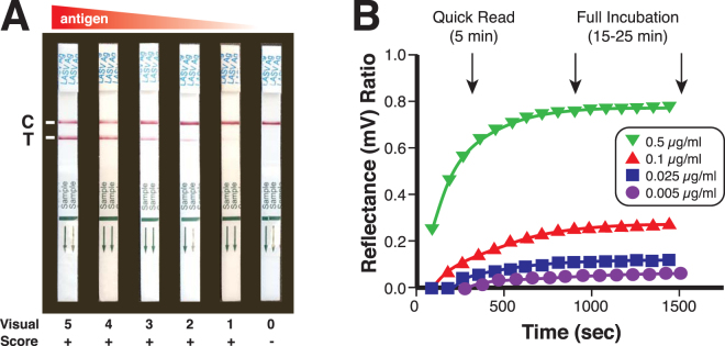
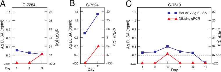
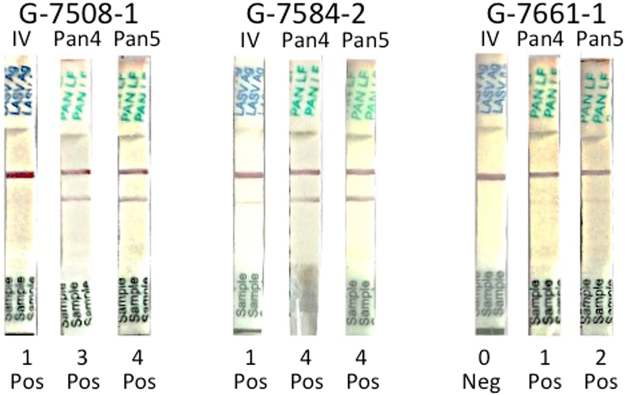
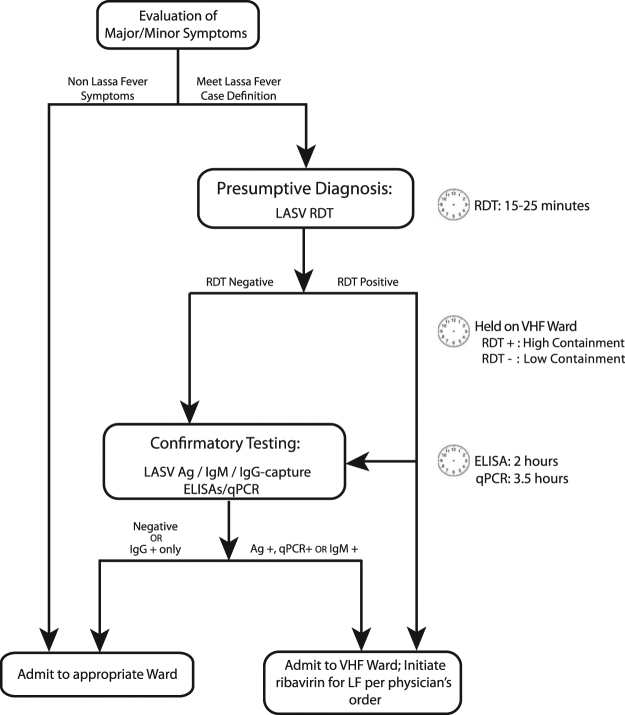
Lassa fever, a hemorrhagic fever caused by Lassa virus (LASV), is endemic in West Africa. It is difficult to distinguish febrile illnesses that are common in West Africa from Lassa fever based solely on a patient's clinical presentation. The field performance of recombinant antigen-based Lassa fever immunoassays was compared to that of quantitative polymerase chain assays (qPCRs) using samples from subjects meeting the case definition of Lassa fever presenting to Kenema Government Hospital in Sierra Leone. The recombinant Lassa virus (ReLASV) enzyme-linked immunosorbant assay (ELISA) for detection of viral antigen in blood performed with 95% sensitivity and 97% specificity using a diagnostic standard that combined results of the immunoassays and qPCR. The ReLASV rapid diagnostic test (RDT), a lateral flow immunoassay based on paired monoclonal antibodies to the Josiah strain of LASV (lineage IV), performed with 90% sensitivity and 100% specificity. ReLASV immunoassays performed better than the most robust qPCR currently available, which had 82% sensitivity and 95% specificity. The performance characteristics of recombinant antigen-based Lassa virus immunoassays indicate that they can aid in the diagnosis of LASV Infection and inform the clinical management of Lassa fever patients.


Conflict of interest statement

The Viral Hemorrhagic Fever Consortium (available at: http://www.vhfc.org) is a partnership of academic and industry scientists who are developing diagnostic tests, therapeutic agents, and vaccines for Lassa fever, Ebola, and other severe diseases. Tulane University and its various academic and industry partners have filed US and foreign patent applications on behalf of the consortium for several of these technologies. Technical information may also be kept as trade secrets. If commercial products are developed, consortium members may receive royalties or profits. This does not alter our adherence to all policies of the NIH and Scientific Reports on sharing data and materials. All authors have submitted the ICMJE Form for Disclosure of Potential Conflicts of Interest. Conflicts that the editors consider relevant to the content of the manuscript have been disclosed.

Figures

Figure 1
Figure 1
Development of a recombinant antigen LASV lateral flow immunoassay. (Panel A) The ReLASV RDT is designed as a dipstick style lateral flow immunoassay. It can be visually scored on a scale of 0 to 5. (Panel B) Scanning densitometry can quantify ReLASV RDT signal kinetics (mean of 3 scans at 90 sec intervals, ratio = test line ÷ control line). NP Ag concentrations are provided in the inset.
Figure 2
Figure 2
Clinical effectiveness of ReLASV RDT, and Ag, IgM, and IgG ELISAs. Comparisons between suspected Lassa fever cases and Sierra Leonean and United States control groups revealed significant differences in ELISA OD or mean visual score. (Panel A) Ag ELISA. (Panel B) RDT. (Panel C) IgM ELISA. (Panel D) IgG ELISA.
Figure 3
Figure 3
Correlations between quantitative values for quantitative polymerase chain reaction assays and ReLASV immunoassay. Cycle threshold (Ct) values, which are inversely related to viral genome equivalents, obtained by Nikisins or Trombley qPCR were compared (Panel A). Densitometry scan ratios between the test and control bands of ReLASV RDT and the amount of LASV antigen (µg/ml) detected by ReLASV (Panel B). Nikisins qPCR values were compared to the quantitative vales of the ReLASV ELISA (Panel C) or RDT (Panel C). Only concordant results were compared. R-squares (R2) values for fit to the linear regression line are indicated.
Figure 4
Figure 4
Earlier detection of Lassa virus in blood with ReLASV immunoassays than with quantitative polymerase chain reaction assays. Comparison of quantitative values for ReLASV Ag ELISA (µg/ml) and Nikisins qPCR (cycle threshold, Ct) in samples that were positive on ReLASV Ag ELISA and RDT on day of presentation (day 1), but negative on Nikisins qPCR are shown (panel A: G-7284, panel B: G-7524, panel C: G-7619). Blue squares: ReLASV Ag levels (µg/ml). Red triangles: Nikisins qPCR (Ct). Dashed line: Nikisins qPCR cycle threshold (Ct) cutoff = 37 cycles).
Figure 5
Figure 5
Clinical chemistry results for patients whose samples were antigen and RDT positive, but qPCR negative. Samples that were Antigen and RDT positive and negative on one or both qPCR assays with sufficient sample volume were compared using a Piccolo Clinical Chemistry Analyzer. Levels of blood creatinine (CRE), alkaline phosphatase (ALP), alanine transaminase (ALT), aspartate aminotransferase (AST). Total bilirubin (tBIL) and albumin are compared. Blue shading indicates normal ranges for the various measures. The T bars indicate standard errors.
Figure 6
Figure 6
Increased sensitivity of prototype Pan-Lassa RDT over Josiah (lineage IV) RDT. Selected samples that were weak (G-7508-1, G-7584-2) or negative (G-7661-1) on the ReLASV lineage IV RDT were run on Pan Lassa RDT prototypes (Pan4 = lot 4, Pan 5 = lot 5).
Figure 7
Figure 7
Proposed diagnostic algorithm for Lassa fever. Use of recombinant Lassa immunodiagnostics for presumptive diagnosis of patients with suspected Lassa fever at KGH and selected PHUs in Sierra Leone.

References

    1. Shaffer JG, et al. Lassa fever in post-conflict Sierra Leone. PLoS neglected tropical diseases. 2014;8:e2748. doi: 10.1371/journal.pntd.0002748. - DOI - PMC - PubMed
    1. Andersen KG, et al. Clinical Sequencing Uncovers Origins and Evolution of Lassa Virus. Cell. 2015;162:738–750. doi: 10.1016/j.cell.2015.07.020. - DOI - PMC - PubMed
    1. WHO. Lassa Fever – Benin, Togo and Burkina Faso. Disease outbreak news 10 March 2017 (2017).
    1. Manning JT, Forrester N, Paessler S. Lassa virus isolates from Mali and the Ivory Coast represent an emerging fifth lineage. Frontiers in microbiology. 2015;6:1037. doi: 10.3389/fmicb.2015.01037. - DOI - PMC - PubMed
    1. ECDC. Lassa fever in Nigeria, Benin, Togo, Germany and USA. European Centre for Disease Prevention and Control 23 March 2016 (2016).

Publication types

LinkOut - more resources