Case Reports
. 2018 Jan 31:2018:9840405.
doi: 10.1155/2018/9840405. eCollection 2018.

A Rare Case of Systemic AL Amyloidosis with Muscle Involvement: A Misleading Diagnosis

Affiliations
Case Reports

A Rare Case of Systemic AL Amyloidosis with Muscle Involvement: A Misleading Diagnosis

Fabrizio Accardi et al. Case Rep Hematol. .

Abstract

Muscle involvement in AL amyloidosis is a rare condition, and the diagnosis of amyloid myopathy is often delayed and underdiagnosed. Amyloid myopathy may be the initial manifestation and may precede the diagnosis of systemic AL amyloidosis. Here, we report the case of a 73-year-old man who was referred to our center for a monoclonal gammopathy of undetermined significance (MGUS) diagnosed since 1999. He reported a progressive weakness of proximal muscles of the legs with onset six months previously. Muscle biopsy showed mild histopathology featuring alterations of nonspecific type with a mixed myopathic and neurogenic involvement, and the diagnostic turning point was the demonstration of characteristic green birefringence under cross-polarized light following Congo red staining of perimysial vessels. Transmission electron microscopy (TEM) confirmed amyloid fibrils around perimysial vessels associated with collagen fibrils. A stepwise approach to diagnosis and staging of this disorder is critical and involves confirmation of amyloid deposition, identification of the fibril type, assessment of underlying amyloidogenic disorder, and evaluation of the extent and severity of amyloidotic organ involvement.


Figures
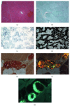
Figure 1
Figure 1
Muscle biopsy general features and the presence of amyloid substance. (a) Hematoxylin and eosin staining: the muscle cells show regular morphological characteristics with the exception of one hypercontracted cell; (b) Gomori staining: normal histological picture; (c) succinate dehydrogenase staining (SDH): the staining shows mild changes in myofibrillar texture; (d) ATPase pH 9.4: normal muscle fiber typing and distribution. Congo red staining without (e) or with polarized light (f): presence of amyloid around/inside the wall of perimysial vessels (×20); (g) Thioflavin S staining with fluorescence microscope: amyloid substance appears bright green in dark field. It completely includes the vessel wall of some perimysial arterioles (×20).
Figure 2
Figure 2
Presence of amyloid fibrils confirmed by electron microscopy. Muscle biopsy was routinely fixed in 2.5% glutaraldehyde in cacodylate buffer, postfixed in osmium tetroxide, and dehydrated and embedded in Araldite; thin sections were studied under Philips CM100 TEM fibrillary structures likely resembling amyloid fibrils in the perimysial pericapillary area (a) (bar = 0.1 µm); an endomysial capillary lumen with a lightly thicker wall without amyloid fibrils (b) (bar = 0.2 µm). Postembedding immunostaining with a polyclonal anti-kappa light chain antibody (Dako, 1 : 100) thin section was studied under a Jeol JEM-1400 Plus electron microscope (c).
Figure 3
Figure 3
Presence of amyloid in the salivary gland. (a) Hematoxylin and eosin staining: some more dark areas between the muscle fibers are observed (×10). Olympus Bx51 optical microscope. (b) Congo red staining with polarized light: apple green birefringence. (c) Postembedding immunostaining: amyloid fibrils are intensely and specifically immunostained with anti-kappa light chain antibody (Dako, 1 : 100). Jeol JEM-1400 Plus electron microscope.

References

    1. Merlini G., Wechalekar A. D., Palladini G. Systemic light chain amyloidosis: an update for treating physicians. Blood. 2013;121(26):5124–5130. doi: 10.1182/blood-2013-01-453001. - DOI - PubMed
    1. Muchtar E., Derudas D., Mauermann M., et al. Systemic immunoglobulin light chain amyloidosis–associated myopathy: presentation, diagnostic pitfalls, and outcome. Mayo Clinic Proceedings. 2016;91(10):1354–1361. doi: 10.1016/j.mayocp.2016.06.027. - DOI - PubMed
    1. Liewluck T., Milone M. Characterization of isolated amyloid myopathy. European Journal of Neurology. 2017;24(12):1437–1445. doi: 10.1111/ene.13448. - DOI - PubMed
    1. Lousada I., Comenzo R. L., Landau H., Guthrie S., Merlini G. Light chain amyloidosis: patient experience survey from the Amyloidosis Research Consortium. Advances in Therapy. 2015;32(10):920–928. doi: 10.1007/s12325-015-0250-0. - DOI - PMC - PubMed
    1. Merlini G., Bellotti V. Molecular mechanisms of amyloidosis. New England Journal of Medicine. 2003;349(6):583–596. doi: 10.1056/nejmra023144. - DOI - PubMed

Publication types

LinkOut - more resources