. 2018 Jan;7(1):3-11.

Tissue Engineering in Maxillary Bone Defects

Affiliations

Tissue Engineering in Maxillary Bone Defects

Azizollah Khodakaram-Tafti et al. World J Plast Surg. 2018 Jan.

Abstract

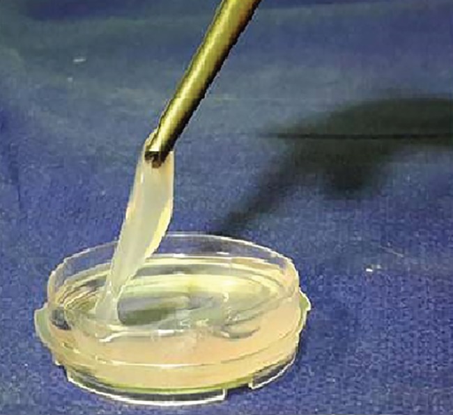
Background: Restoration of craniofacial bone defects has been a concern for oral and maxillofacial surgeons. In this study, the healing effect of fibrin glue scaffold was compared with autologous bone graft in mandibular defects of rabbit.

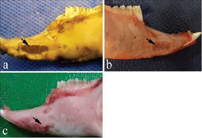
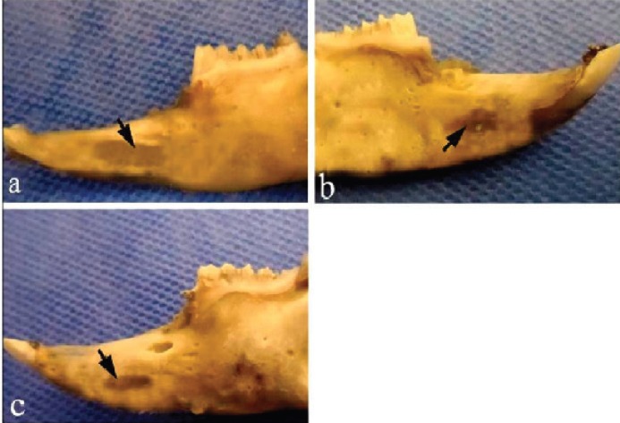
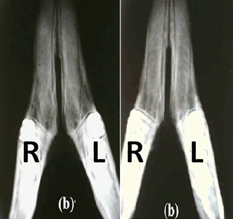
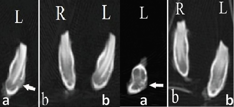
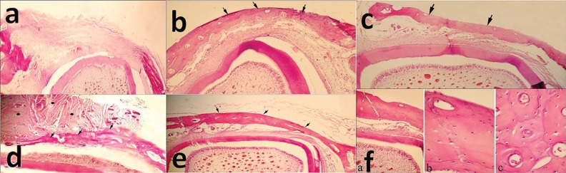
Methods: Bilateral unicortical osteotomy was performed in the diastema region of 10 male Dutch rabbits. The subjects were randomly divided into 2 equal groups. The mandibular defect on the right side was treated with fibrin glue scaffold and the defect on the left side with autologous bone graft provided from iliac crest. After 4 and 8 weeks, five rabbits from each group were sacrificed and the defects were evaluated morphologically, by coronal computed tomography scanning (CT-scan) and by histological examinations.

Results: The healing effect of fibrin glue scaffold and autologous bone graft was similar with appropriate osteogenesis in comparison to the control group.

Conclusion: Using fibrin glue can be a non-invasive treatment of choice in mandibular defects and maxillofacial surgeries when compared with autologous bone graft.

Keywords: Autologous bone graft; Fibrin glue; Mandibular defect; Rabbit; Scaffold.


Conflict of interest statement

The authors declare no conflict of interest.

Figures

Fig. 1
Fig. 1
A gel-like fibrin glue formation after mixing of fibrinogen and thrombin.
Fig. 2
Fig. 2
Macroscopic evaluation of the bone samples 4 weeks after surgery. a) the control defect, b) the defect was filled by fibrin glue alone, c) the defect was filled by autologous bone graft.
Fig. 3
Fig. 3
Macroscopic evaluation of the bone samples 8 weeks after surgery. a) the control defect, b) the defect was filled by fibrin glue alone, c) the defect was filled by autologous bone graft. Arrows denote the defect area.
Fig. 4
Fig. 4
Radiographic observation on day of injury. a) Left R: the defect filled with fibrin glue; Left L: the control defect, b) Right R: the defect filled with fibrin glue; Right L: the defect filled with autologous bone graft after 4 weeks.
Fig. 5
Fig. 5
Coronal CT scanning observation after 4 weeks. Left aL: the control defect, Left bR: the defect filled with fibrin glue; Left bL: the defect filled with autologous bone graft. Coronal CT scanning observation 8 weeks after surgery. Right aL: the control defect, Right bR: the defect filled with fibrin glue; Right bL: the defect filled with autologous bone graft.
Fig. 6
Fig. 6
Histopathological evaluation of the bone samples 4 weeks after surgery: a) the control defect; x60), b) the defect was filled by fibrin glue alone; x60, c) the defect was filled by autologous bone graft; x150. At the 8th week post-surgery: d) formation of a thin cortical bone bridge in control group, while fibrous-immature bone tissue is still presence; x60, e) A complete cortical bone formation that is thicker than 4 weeks post-treatment in fibrin glue group; x60, f) Lamellar bone with less Haversian system in autologous bone graft group; f-a) x150, f-b) x600, f-c) x600 (H&E).

References

    1. Arrigoni E, Lopa S, de Girolamo L, Stanco D, Brini AT. Isolation, characterization and osteogenic differentiation of adipose-derived stem cells: from small to large animal models. Cell Tissue Res. 2009;338:401–11. - PubMed
    1. Moore WR, Graves SE, Bain GI. Synthetic bone graft substitutes. ANZ J Surg. 2001;71:354–61. - PubMed
    1. Mehrabani D M, Babazadeh M, Tanideh N, Zare S, Hoseinzadeh S, Torabinejad S et al. The healing effect of adipose-derived mesenchymal stem cells in full-thickness femoral articular cartilage defects of rabbit. Int J Organ Transplant Med. 2015;6:165–75. - PMC - PubMed
    1. Tanideh N, Bagheri MH, Dehghani Nazhvani S, Nikahval B, Mojtahed Jaberi F, Mehrabani D. MRI and CT evaluations of an invented bioglue in experimentally induced articular cartilage defects in rabbits. Comp Clin Pathol. 2014;23:1545–50.
    1. Tanideh N, Dehghani Nazhvani S, Mojtahed Jaberi F, Mehrabani D, Rezazadeh S, Pakbaz S et al. The healing effect of bioglue in experimental rabbit model of femoral condyle articular cartilage defect. Iran Red Crescent Med J. 2011;13:544–9. - PMC - PubMed

LinkOut - more resources