

Analysis of microRNAs Expression Profiles in Madin-Darby Bovine Kidney Cells Infected With Caprine Parainfluenza Virus Type 3
- PMID: 29651410
- PMCID: PMC5885596
- DOI: 10.3389/fcimb.2018.00093
Analysis of microRNAs Expression Profiles in Madin-Darby Bovine Kidney Cells Infected With Caprine Parainfluenza Virus Type 3
Abstract
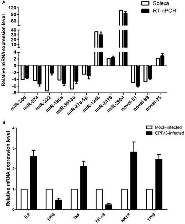
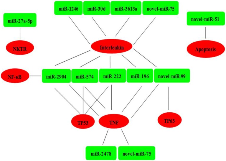
Caprine parainfluenza virus type 3 (CPIV3) is a newly emerging pathogenic respiratory agent infecting both young and adult goats, and it was identified in eastern China in 2013. Cellular microRNAs (miRNAs) have been reported to be important modulators of the intricate virus-host interactions. In order to elucidate the role of miRNAs in madin-darby bovine kidney (MDBK) cells during CPIV3 infection. In this study, we performed high-throughput sequencing technology to analyze small RNA libraries in CPIV3-infected and mock-infected MDBK cells. The results showed that a total of 249 known and 152 novel candidate miRNAs were differentially expressed in MDBK cells after CPIV3 infection, and 22,981 and 22,572 target genes were predicted, respectively. In addition, RT-qPCR assay was used to further confirm the expression patterns of 13 of these differentially expressed miRNAs and their mRNA targets. Functional annotation analysis showed these up- and downregulated target genes were mainly involved in MAPK signaling pathway, Jak-STAT signaling pathway, Toll-like receptor signaling pathway, p53 signaling pathway, focal adhesion, NF-kappa B signaling pathway, and apoptosis, et al. To our knowledge, this is the first report of the comparative expression of miRNAs in MDBK cells after CPIV3 infection. Our finding provides information concerning miRNAs expression profile in response to CPIV3 infection, and offers clues for identifying potential candidates for antiviral therapies against CPIV3.
Keywords: caprine parainfluenza virus type 3; high-throughput sequencing; host-pathogen interactions; madin-darby bovine kidney cell line; microRNAs.
Figures
Similar articles
-
Bta-miR-98 Suppresses Replication of Caprine Parainfluenza Virus Type 3 Through Inhibiting Apoptosis by Targeting Caspase-3.Front Immunol. 2020 Aug 28;11:1575. doi: 10.3389/fimmu.2020.01575. eCollection 2020. Front Immunol. 2020. PMID: 32983081 Free PMC article.
-
Proteomics analysis reveals heat shock proteins involved in caprine parainfluenza virus type 3 infection.BMC Vet Res. 2019 May 17;15(1):151. doi: 10.1186/s12917-019-1897-6. BMC Vet Res. 2019. PMID: 31101113 Free PMC article.
-
Exosomes promote caprine parainfluenza virus type 3 infection by inhibiting autophagy.J Gen Virol. 2020 Jul;101(7):717-734. doi: 10.1099/jgv.0.001424. J Gen Virol. 2020. PMID: 32427096
-
Interferon-stimulated genes inhibit caprine parainfluenza virus type 3 replication in Madin-Darby bovine kidney cells.Vet Microbiol. 2020 Feb;241:108573. doi: 10.1016/j.vetmic.2019.108573. Epub 2019 Dec 31. Vet Microbiol. 2020. PMID: 31928705
-
The replicative complex of paramyxoviruses: structure and function.Adv Virus Res. 1998;50:101-39. doi: 10.1016/s0065-3527(08)60807-6. Adv Virus Res. 1998. PMID: 9520998 Review. No abstract available.
Cited by
-
Establishment of a cloning-free CRISPR/Cas9 protocol to generate large deletions in the bovine MDBK cell line.J Appl Genet. 2024 May;65(2):399-402. doi: 10.1007/s13353-024-00846-3. Epub 2024 Feb 28. J Appl Genet. 2024. PMID: 38418802 Free PMC article.
-
Analysis of the microRNA Expression Profile of Bovine Monocyte-derived Macrophages Infected with Mycobacterium avium subsp. Paratuberculosis Reveals that miR-150 Suppresses Cell Apoptosis by Targeting PDCD4.Int J Mol Sci. 2019 Jun 1;20(11):2708. doi: 10.3390/ijms20112708. Int J Mol Sci. 2019. PMID: 31159463 Free PMC article.
-
Integrated Analysis of Transcriptome Profiles and lncRNA-miRNA-mRNA Competing Endogenous RNA Regulatory Network to Identify Biological Functional Effects of Genes and Pathways Associated with Johne's Disease in Dairy Cattle.Noncoding RNA. 2024 Jun 28;10(4):38. doi: 10.3390/ncrna10040038. Noncoding RNA. 2024. PMID: 39051372 Free PMC article.
-
Bta-miR-98 Suppresses Replication of Caprine Parainfluenza Virus Type 3 Through Inhibiting Apoptosis by Targeting Caspase-3.Front Immunol. 2020 Aug 28;11:1575. doi: 10.3389/fimmu.2020.01575. eCollection 2020. Front Immunol. 2020. PMID: 32983081 Free PMC article.
-
RNA-Seq and miRNA-Seq data from Epstein-Barr virus-infected tree shrews reveal a ceRNA network contributing to immune microenvironment regulation.Virulence. 2024 Dec;15(1):2306795. doi: 10.1080/21505594.2024.2306795. Epub 2024 Jan 29. Virulence. 2024. PMID: 38251668 Free PMC article.
References
Publication types
MeSH terms
Substances
LinkOut - more resources
Full Text Sources
Other Literature Sources
Research Materials
Miscellaneous