Review
. 2018 Feb 5:2018:2584243.
doi: 10.1155/2018/2584243. eCollection 2018.

Mast Cell, the Neglected Member of the Tumor Microenvironment: Role in Breast Cancer

Affiliations
Review

Mast Cell, the Neglected Member of the Tumor Microenvironment: Role in Breast Cancer

Angélica Aponte-López et al. J Immunol Res. .

Abstract

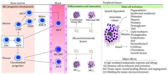
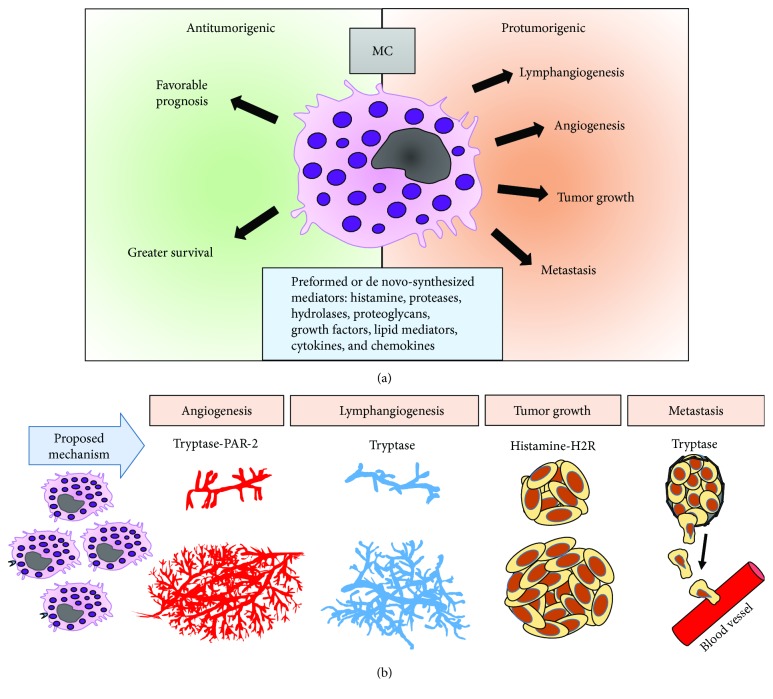
Mast cells are unique tissue-resident immune cells that secrete a diverse array of biologically active compounds that can stimulate, modulate, or suppress the immune response. Although mounting evidence supports that mast cells are consistently infiltrating tumors, their role as either a driving or an opposite force for cancer progression is still controversial. Particularly, in breast cancer, their function is still under discussion. While some studies have shown a protective role, recent evidence indicates that mast cells enhance blood and lymphatic vessel formation. Interestingly, one of the most important components of the mast cell cargo, the serine protease tryptase, is a potent angiogenic factor, and elevated serum tryptase levels correlate with bad prognosis in breast cancer patients. Likewise, histamine is known to induce tumor cell proliferation and tumor growth. In agreement, mast cell depletion reduces the size of mammary tumors and metastasis in murine models that spontaneously develop breast cancer. In this review, we will discuss the evidence supporting protumoral and antitumoral roles of mast cells, emphasizing recent findings placing mast cells as important drivers of tumor progression, as well as the potential use of these cells or their mediators as therapeutic targets.


Figures

Figure 1
Figure 1
Overview of mast cell development, heterogeneity, and activation. Mast cells arise in the bone marrow from hematopoietic stem cells (HSC) via a multipotent progenitor (MPP), which can become a mast cell-committed progenitor (MCP) that exits the bone marrow and migrates to peripheral tissues to complete maturation. Several pathways have been described for murine and human mast cell origin. In mice, MCPs may be derived directly from MPPs or from common myeloid progenitors (CMP). Mast cells may also be derived from the granulocyte/monocyte progenitor (GMP) via an intermediate progenitor (BMCP), identified only in the spleen of C57BL6 mice, which gives rise to basophils and mast cells. In humans, it has been postulated that mast cells originate from a yet unidentified uncommitted progenitor that gives rise to a mast cell/monocyte-committed progenitor (MC/MP) in bone marrow. Alternatively, an MCP population that gives rise exclusively to mast cells has recently been identified in blood. Final differentiation occurs in peripheral tissues, where microenvironmental factors determine the phenotype of the mature mast cells. Mast cells exhibit marked phenotypic and functional heterogeneity. Two major subtypes have been described in both rodents and humans, in the former as mucosal and connective tissue mast cells and in the latter as tryptase- and chymase-rich mast cells (MCTC) and those that mainly contain tryptase (MCT). The right end diagram illustrates the distinct classes of bioactive molecules and their temporality of release upon mast cell activation in tissues. See text for a more detailed explanation.
Figure 2
Figure 2
Role of mast cells in breast cancer. (a) The influence of mast cells in breast cancer prognosis is still a matter of discussion. Mast cells contain a great variety of bioactive components that may exert both pro- and antitumor effects. On the one hand, in vitro and in vivo studies support that mast cells exhibit protumor activity through promotion of lymphatic and blood vessel formation, tumor growth, and metastasis (orange right side). On the other hand, several population studies also associate mast cells with a greater survival and favorable prognosis (green left side). (b) Some bioactive molecules of mast cells documented to have protumorigenic effects are tryptase through its receptor PAR-2 and histamine through H2 receptor. The cancer processes in which these compounds have been associated are indicated.

References

    1. Bermejo-Martin J. F., Martin-Loeches I., Bosinger S. Inflammation and infection in critical care medicine. Mediators of Inflammation. 2014;2014:2. doi: 10.1155/2014/456256.456256 - DOI - PMC - PubMed
    1. Medzhitov R. Inflammation 2010: new adventures of an old flame. Cell. 2010;140(6):771–776. doi: 10.1016/j.cell.2010.03.006. - DOI - PubMed
    1. Vendramini-Costa D. B., Carvalho J. E. Molecular link mechanisms between inflammation and cancer. Current Pharmaceutical Design. 2012;18(26):3831–3852. doi: 10.2174/138161212802083707. - DOI - PubMed
    1. Virchow R. Cellular pathology as based upon physiological and pathological histology. Philadelphia, PA, USA: J. B. Lippincott and Co.; 1863. - DOI - PubMed
    1. Eiro N., Vizoso F. J. Inflammation and cancer. World Journal of Gastrointestinal Surg. 2012;4(3):62–72. doi: 10.4240/wjgs.v4.i3.62. - DOI - PMC - PubMed