

Outcomes of Intraoperative OCT-Assisted Epiretinal Membrane Surgery from the PIONEER Study
- PMID: 29651467
- PMCID: PMC5891156
- DOI: 10.1016/j.oret.2017.05.006
Outcomes of Intraoperative OCT-Assisted Epiretinal Membrane Surgery from the PIONEER Study
Abstract
Purpose: To assess the retinal architecture changes which occur during epiretinal membrane (ERM) surgery, utilizing intraoperative optical coherence tomography (iOCT).
Design: Prospective multi-surgeon single center study.
Subjects/participants: Subjects from the PIONEER iOCT study who underwent surgical intervention for management of ERM.
Methods: All subjects underwent vitrectomy with ERM peeling with optional internal limiting membrane (ILM) peeling. Preoperative, intraoperative, and postoperative quantitative and qualitative OCT assessments were performed. Clinical characteristics including visual acuity outcomes, central subfield thickness and complications including ERM recurrence and need for reoperation were assessed at 3, 6 and 12 months following surgery for membrane peeling, as available.
Main outcome measures: Visual acuity outcomes, anatomic outcomes and complications including ERM recurrence. Microarchitectural alterations (i.e. retinal layer changes) following membrane peeling visualized with iOCT.
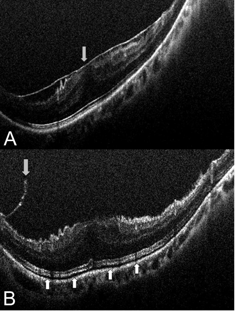
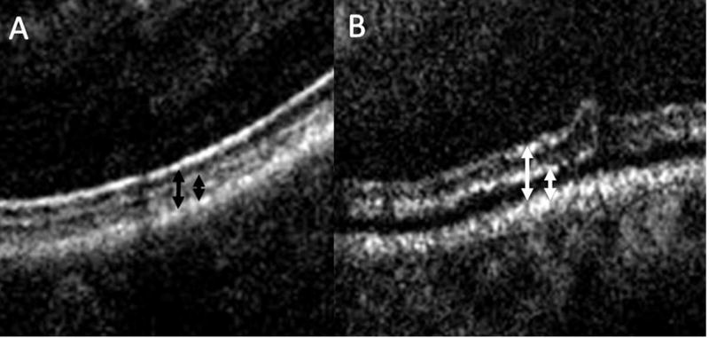
Results: Seventy-six were identified and included in this analysis of clinical outcomes and quantitative OCT assessment. Twenty-four eyes were excluded due to insufficient intraoperative OCT quality for quantitative assessment. The mean preoperative VA measured 20/63. The mean postoperative VA at 3 months was 20/41 (p<0.0001), at 6 months measured 20/36 (p < 0.0001), and at 12 months measured 20/33 (p < 0.0001). Preoperative mean central subfield thickness (CST) was 426 microns. At 3 months, the mean CST improved to 377 microns (p < 0.0001). The 6-month postoperative CST was 367 microns (p < 0.0001) and the 12-month postoperative CST measured 359 microns (p < 0.0001). Immediately following membrane peeling, the distance between the retinal pigment epithelium and the ellipsoid zone as well as the distance between the retinal pigment epithelium and the cone outer segment tips/interdigitation zone significantly increased (p < 0.001). iOCT identified occult residual membranes in 12% of cases and confirmed complete membrane peeling contrary to surgeon impression in 9% of cases. Reoperation was required for recurrent ERM in 1% of eyes.
Conclusions: iOCT-assisted ERM peeling resulted in significant improvement in visual acuity, reduction in macular thickness, and low recurrence rate. Additional research is needed with randomized clinical trials to better define the comparative success rates of image-guided ERM surgery to standard surgical visualization techniques.
Figures
References
-
- Schmidt-Erfurth U, Leitgeb RA, Michels S, et al. Three-dimensional ultrahigh-resolution optical coherence tomography of macular diseases. Invest Ophthalmol Vis Sci. 2005;46(9):3393–402. - PubMed
-
- Chen TC, Cense B, Pierce MC, et al. Spectral domain optical coherence tomography: ultra-high speed, ultra-high resolution ophthalmic imaging. Arch Ophthalmol. 2005;123(12):1715–20. - PubMed
Grants and funding
LinkOut - more resources
Full Text Sources
Other Literature Sources