

Effect of renal sympathetic denervation on ventricular and neural remodeling
- PMID: 29651618
- PMCID: PMC6890580
- DOI: 10.1007/s00059-018-4698-y
Effect of renal sympathetic denervation on ventricular and neural remodeling
Abstract
Background: This study assessed the therapeutic effects of renal sympathetic denervation (RDN) on post-myocardial infarction (MI) ventricular remodeling and sympathetic neural remodeling in dogs. The possible mechanisms and optimal time for treatment are discussed.
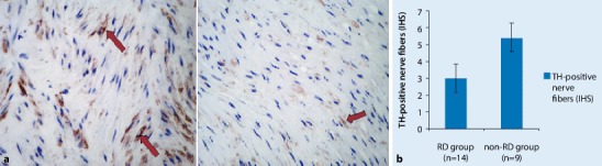
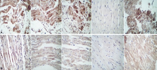
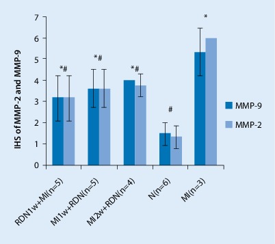
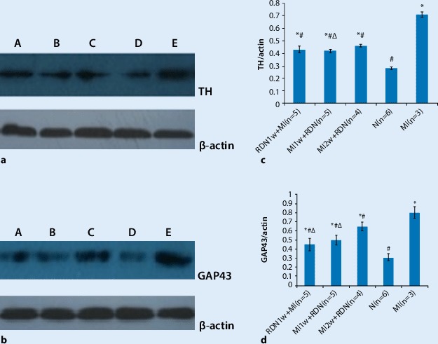
Methods: We randomly assigned 30 dogs to five groups: RDN 1 week before MI (RDN1w + MI; n = 6), RDN 1 week after MI (MI1w + RDN; n = 6), RDN 2 weeks after MI (MI2w + RDN; n = 6), control (N; n = 6), and MI (n = 6). A canine model of myocardial infarction was established by interventional occlusion with a gelatin sponge via the femoral artery. Brain natriuretic peptide (BNP) and endothelin-1 (ET-1) levels were measured and echocardiography was performed to assess cardiac function and heart size. All dogs were killed at the end of the experiment and samples of cardiac and renal arteries were obtained. The expression of matrix metalloproteinase (MMP)-2 and MMP-9 in cardiac and of tyrosine hydroxylase (TH) in renal arteries was assessed by immunohistochemistry. Sympathetic innervations in the infarction border zone were investigated via Western blotting and real-time PCR.
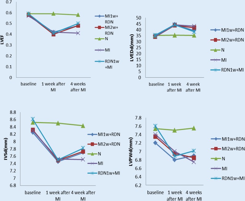
Results: Left ventricular function in the MI group decreased significantly, while plasma BNP and ET-1 levels as well as MMP-2 and MMP-9 expression increased. Compared with the MI group, the RD groups showed significantly reduced MMP‑2, MMP‑9, TH, and growth-associated protein (GAP) 43 expression in the RDN1w + MI, MI1w + RDN, and MI2w + RDN groups was significantly improved. Additionally, the expression of TH in renal arteries decreased after RDN.
Conclusion: RDN has preventive and therapeutic effects on post-MI ventricular remodeling and sympathetic neural remodeling. The mechanism of RDN is likely mediated through restraint of renal sympathetic nerve activity.
Hintergrund: In der vorliegenden Studie wurden die therapeutischen Auswirkungen der renalen sympathischen Denervierung (RDN) auf das ventrikuläre Remodeling nach Myokardinfarkt (MI) und das sympathische neurale Remodeling bei Hunden untersucht. Mögliche Mechanismen und der optimale Zeitpunkt der Behandlung werden erörtert.
Methoden: Randomisiert wurden 30 Hunde auf 5 Gruppen verteilt: RDN 1 Woche vor MI (RDN1w + MI; n = 6), RDN 1 Woche nach MI (MI1w + RDN; n = 6), RDN 2 Wochen nach MI (MI2w + RDN; n = 6), Kontrolle (N, n = 6) und MI (n = 6). Ein Hundemodell des Herzinfarkts wurde durch interventionelle Okklusion mit einem Gelatineschwamm über die A. femoralis etabliert. Die Werte für BNP („brain natriuretic peptide“) und Endothelin-1 (ET-1) wurden ermittelt und eine Echokardiographie zur Beurteilung der kardialen Funktion und Herzgröße durchgeführt. Alle Hunde wurden am Ende des Experiments getötet und Proben aus den Herz- und Nierenarterien gewonnen. Die Expression von MMP-2 und MMP-9 in Herzarterien und von Tyrosinhydroxylase (TH) in den Nierenarterien wurde immunohistochemisch ermittelt. Sympathische Innervationen in der Infarktgrenzzone wurden per Westernblot und Echtzeitpolymerasekettenreaktion („real-time PCR“) untersucht.
Ergebnisse: Die linksventrikuläre Funktion in der MI-Gruppe nahm signifikant ab, während die Werte für BNP und ET-1 im Plasma sowie für die MMP-2- und die MMP-9-Expression erhöht waren. Im Vergleich zur MI-Gruppe war die linksventrikuläre Ejektionsfraktion, MMP‑2-, MMP‑9-, TH- und „growth-associated protein“(GAP)-43-Expression in den Gruppen RDN1w + MI, MI1w + RDN und MI2w + RDN signifikant verbessert. Außerdem nahm die Expression von TH in den Nierenarterien nach RDN ab.
Schlussfolgerung: Die RDN hat präventive und therapeutische Auswirkungen auf das ventrikuläre Remodeling nach MI und auf das sympathische renale Remodeling. Wahrscheinlich wird der Mechanismus der RDN durch eine Begrenzung der Aktivität des renalen N. sympathicus vermittelt.
Keywords: Cardiac remodeling; Experimental animal model; Heart attack; Myocardial infarction; Sympathectomy.
Conflict of interest statement
L. Wang, G. Wei, L. Song, C. Li, F. Zhang, Y. Yang, and C. Lu declare that they have no competing interests.
Figures


References
-
- Xiao-fei W, Ren-fei L, Li-hong B. Establishing a canine model of precise acute myocardial infarction by interventional occlusion with gelatin sponge via femoral artery.progress in modern. Biomedicine (Taipei) 2010;10(19):3601–3605.
-
- Yarbrough WM, Mukherjee R, Escobar GP, Mingoia JT, Sample JA, Hendrick JW, Dowdy KB, McLean JE, Lowry AS, O’Neill TP, Spinale FG. Selective targeting and timing of matrix metalloproteinase inhibition in post-myocardial infarction remodeling. Circulation. 2003;108(14):1753–1759. doi: 10.1161/01.CIR.0000091087.78630.79. - DOI - PubMed
-
- Hayashidani S, Tsutsui H, Ikeuchi M, Shiomi T, Matsusaka H, Kubota T, Imanaka-Yoshida K, Itoh T, Takeshita A. Targeted deletion of MMP-2 attenuates early LV rupture and late remodeling after experimental myocardial infarction. Am J Physiol Heart Circ Physiol. 2003;285(3):H1229–35. doi: 10.1152/ajpheart.00207.2003. - DOI - PubMed
MeSH terms
LinkOut - more resources
Full Text Sources
Other Literature Sources
Medical
Miscellaneous
