. 2019 Jan;44(1):61-77.
doi: 10.1007/s11064-018-2526-7. Epub 2018 Apr 12.

In the Telencephalon, GluN2C NMDA Receptor Subunit mRNA is Predominately Expressed in Glial Cells and GluN2D mRNA in Interneurons

Affiliations

In the Telencephalon, GluN2C NMDA Receptor Subunit mRNA is Predominately Expressed in Glial Cells and GluN2D mRNA in Interneurons

Hassan A Alsaad et al. Neurochem Res. 2019 Jan.

Abstract

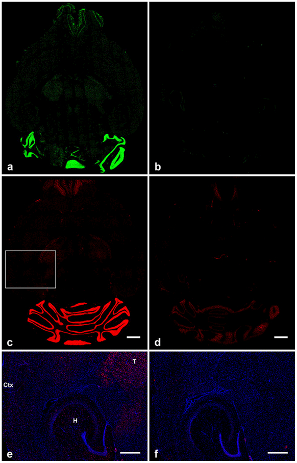
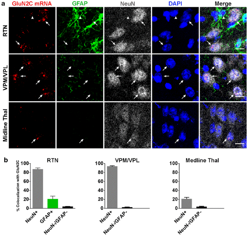
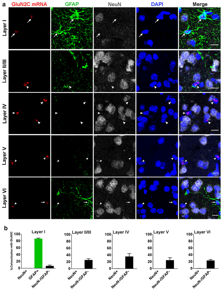
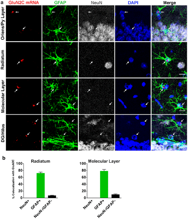
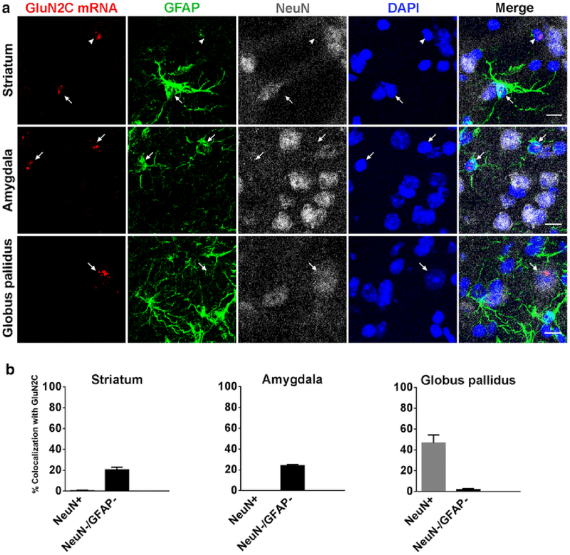
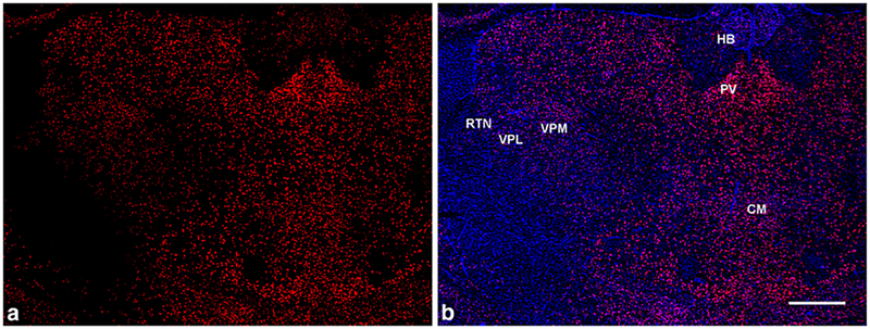
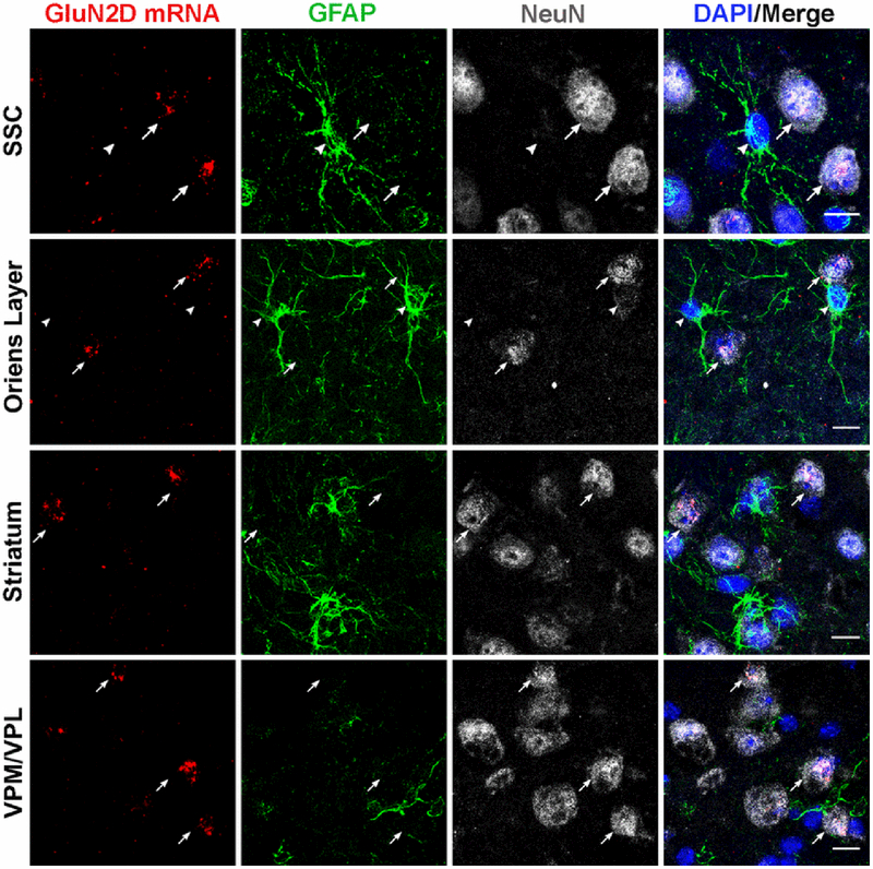
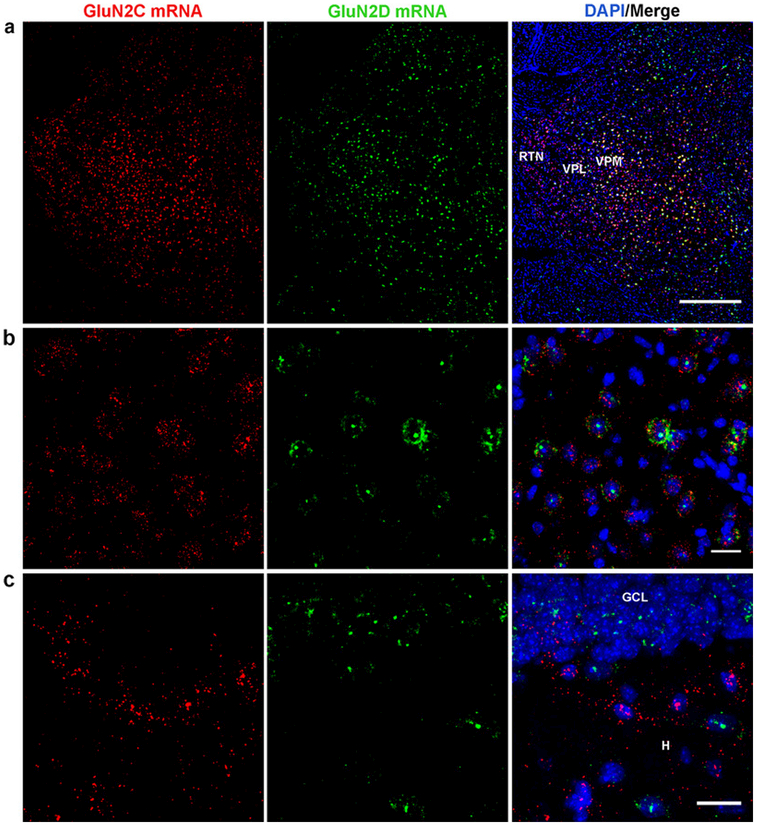
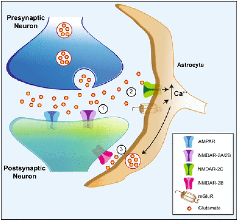
N-methyl-D-aspartate receptors (NMDARs) are widely distributed in the brain with high concentrations in the telencephalon where they modulate synaptic plasticity, working memory, and other functions. While the actions of the predominate GluN2 NMDAR subunits, GluN2A and GluN2B are relatively well understood, the function of GluN2C and GluN2D subunits in the telencephalon is largely unknown. To better understand the possible role of GluN2C subunits, we used fluorescence in situ hybridization (FISH) together with multiple cell markers to define the distribution and type of cells expressing GluN2C mRNA. Using a GluN2C-KO mouse as a negative control, GluN2C mRNA expression was only found in non-neuronal cells (NeuN-negative cells) in the hippocampus, striatum, amygdala, and cerebral cortex. For these regions, a significant fraction of GFAP-positive cells also expressed GluN2C mRNA. Overall, for the telencephalon, the globus pallidus and olfactory bulb were the only regions where GluN2C was expressed in neurons. In contrast to GluN2C, GluN2D subunit mRNA colocalized with neuronal and not astrocyte markers or GluN2C mRNA in the telencephalon (except for the globus pallidus). GluN2C mRNA did, however, colocalize with GluN2D in the thalamus where neuronal GluN2C expression is found. These findings strongly suggest that GluN2C has a very distinct function in the telencephalon compared to its role in other brain regions and compared to other GluN2-containing NMDARs. NMDARs containing GluN2C may have a specific role in regulating L-glutamate or D-serine release from astrocytes in response to L-glutamate spillover from synaptic activity.

Keywords: Astrocyte; Cortex; GluN2C; GluN2D; Hippocampus; L-glutamate; N-methyl-D-aspartate receptor; mRNA.


Figures

Fig. 1
Fig. 1
Overall distribution and specificity of fluorescein-tagged (a, b), and digoxigenin-tagged (c-f) antisense RNA probe binding in horizontal mouse brain sections. (a, c) GluN2C mRNA signal in wild-type mouse brain showing the highest expression levels in the granular layer of the cerebellum. b, d, GluN2C-KO mouse brain sections showing very low (b) and low (d) background staining in cerebellum and olfactory bulb that was independent of the tag used. (e, f) Enlarged images of the cortex, hippocampus, and thalamus which are highlighted by the box in (c). Scattered GluN2C signal is displayed in these regions of wild-type mouse (e) but not in GluN2C-KO mouse (f). T, thalamus; Ctx, cortex; H, hippocampus. Scale bars: c, d, 1 mm; e, f, 500 μm
Fig. 2
Fig. 2
Expression and colocalization of GluN2C mRNA in thalamus. (a) GluN2C mRNA colocalizes with NeuN-positive cells in RTN, VPM/VPL, and midline thal (arrows). GluN2C mRNA is expressed by a GFAP-positive cell in the RTN (arrowhead). (b) Percent of NeuN-positive, GFAP-positive, or NeuN-negative/GFAP-negative cells that were also labeled by GluN2C probe in the three thalamic regions. Scale bar = 10μm
Fig. 3
Fig. 3
GluN2C mRNA expression in multiple layers of retrosplenial cortex. (a) GluN2C mRNA is expressed in GFAP-positive cells in layer I (arrows). GluN2C mRNA (arrows/arrowheads) does not colocalize with NeuN-positive cells in any layer of the RSC. GluN2C mRNA colocalizes with NeuN-negative/GFAP-negative cells in the deep layers of the RSC. (b) Percent of NeuN-positive, GFAP-positive, or NeuN-negative/GFAP-negative cells that were also labeled by GluN2C probe in different layers of RSC. Scale bar = 10μm
Fig. 4
Fig. 4
GluN2C mRNA expression in somatosensory cortex. (a) GluN2C mRNA is expressed by GFAP-positive cells in layer I and layer VI (arrows). GluN2C mRNA does not colocalize with NeuN-positive cells in any layers of the SSC (arrows/arrowheads). GluN2C mRNA colocalizes with NeuN-negative/GFAP-negative cells (arrowheads). (b) Percent of NeuN-positive, GFAP-positive, or NeuN-negative/GFAP-negative cells that were also labeled by GluN2C probe in each of the layers of SSC. Scale bar = 10μm
Fig. 5
Fig. 5
GluN2C mRNA expression in hippocampus. (a) GluN2C mRNA colocalizes with GFAP-positive cells in stratum oriens, the pyramidal cell layer, stratum radiatum, the molecular layer, the dentate gyrus-granular cell layer, and the hilus (arrows). GluN2C mRNA does not colocalize with NeuN-positive cells in these regions. GluN2C mRNA colocalizes with NeuN-negative/GFAP-negative cells. (b) Percent of NeuN-positive, GFAP-positive, or NeuN-negative/GFAP-negative cells that were also labeled by GluN2C probe in the stratum radiatum and the molecular layer. Scale bar = 10μm
Fig. 6
Fig. 6
GluN2C mRNA expression in striatum, amygdala, and globus pallidus. (a) GluN2C mRNA colocalizes with GFAP-positive cells in the striatum and the amygdala (arrows). GluN2C mRNA colocalizes with NeuN-positive cells in the globus pallidus (arrow). GluN2C mRNA is also found in NeuN-negative/GFAP-negative cells (arrowhead). (b) Percent of NeuN-positive and NeuN-negative/GFAP-negative cells that were also labeled by GluN2C probe in the striatum, amygdala, and globus pallidus. Scale bar = 10μm
Fig. 7
Fig. 7
GluN2D mRNA distribution in the thalamus in coronal brain sections. (a, b) GluN2D signal (red) is higher in the midline thalamic nuclei (including paraventricular (PV) and central medial (CM) nuclei) compared to the habenula (HB) and the lateral thalamus - ventrobasal complex (VPM/VPL) and RTN. (b) GluN2D mRNA probe (red) co-stained with DAPI (blue). Scale bar = 500μm
Fig. 8
Fig. 8
GluN2D mRNA expression colocalizes with NeuN in the somatosensory cortex, hippocampus (e.g. stratum oriens), striatum and the thalamic VPM/VPL nuclei (arrows). GluN2D mRNA did not colocalize with GFAP-positive cells (arrowheads). Scale bar = 10μm
Fig. 9
Fig. 9
Localization of GluN2C and GluN2D mRNA in hippocampus and thalamus. GluN2C and GluN2D mRNA display partial colocalization in the lateral thalamus (VPM/VPL,RTN) low magnification (a) and high magnification (VPM/VPL) (b). (c) GluN2C and GluN2D mRNA did not colocalize in the dentate gyrus granule cell layer (GCL) or hilus (H). (c). Scale bars: a, 500 μm; b, c, 20 μm
Fig. 10
Fig. 10
Schematic diagram showing a potential role for GluN2C-containing NMDARs in the tripartite synapse. (1) L-Glutamate released from presynaptic nerve endings activates AMPA receptors and GluN2A-and/or GluN2B-containing NMDARs (NMDAR-2A/2B) on the postsynaptic dendrite. (2) With sufficient synaptic activation, glutamate spillover activates GluN2C-containing NMDARs (NMDAR-2C) and metabotropic glutamate receptors (mGluR) on the astrocyte, leading to increased cytoplasmic calcium levels from extracellular and intracellular sources, respectively. (3) In response to elevated intracellullar calcium from intracellular stores, and possibly from GluN2C currents, astrocytes release glutamate (and other gliotransmitters) which acts on extrasynaptic GluN2B-containing NMDARs (NMDAR-2B) on the postsynaptic structure on the same neuron, and potentially additional neurons, to modulate excitability and synaptic plasticity.

References

    1. Yamamoto H, Hagino Y, Kasai S and Ikeda K, (2015) Specific Roles of NMDA Receptor Subunits in Mental Disorders. Curr. Mol. Med 15, 193–205. - PMC - PubMed
    1. Traynelis SF, Wollmuth LP, McBain CJ, Menniti FS, Vance KM, Ogden KK, Hansen KB, Yuan H, Myers SJ and Dingledine R, (2010) Glutamate receptor ion channels: structure, regulation, and function. Pharmacol. Rev 62, 405–496. - PMC - PubMed
    1. Buller AL, Larson HC, Schneider BE, Beaton JA, Morrisett RA and Monaghan DT, (1994) The molecular basis of NMDA receptor subtypes: native receptor diversity is predicted by subunit composition. J. Neurosci 14, 5471–5484. - PMC - PubMed
    1. Monyer H, Burnashev N, Laurie DJ, Sakmann B and Seeburg PH, (1994) Developmental and regional expression in the rat brain and functional properties of four NMDA receptors. Neuron. 12, 529–540. - PubMed
    1. Mishina M, Mori H, Araki K, Kushiya E, Meguro H, Kutsuwada T, Kashiwabuchi N, Ikeda K, Nagasawa M and Yamazaki M, (1993) Molecular and functional diversity of the NMDA receptor channel. Ann. N. Y. Acad. Sci 707, 136–152. - PubMed

LinkOut - more resources