Review
. 2018;16(9):1385-1395.
doi: 10.2174/1570159X16666180412110919.

Inflammatory Events Following Subarachnoid Hemorrhage (SAH)

Affiliations
Review

Inflammatory Events Following Subarachnoid Hemorrhage (SAH)

U C Schneider et al. Curr Neuropharmacol. 2018.

Abstract

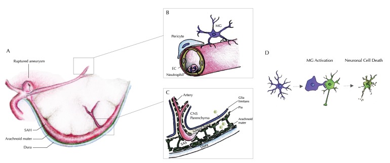
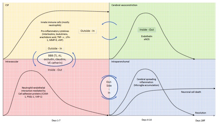
Acute SAH from a ruptured intracranial aneurysm contributes for 30% of all hemorrhagic strokes. The bleeding itself occurs in the subarachnoid space. Nevertheless, injury to the brain parenchyma occurs as a consequence of the bleeding, directly, via several well-defined mechanisms and pathways, but also indirectly, or secondarily. This secondary brain injury following SAH has a variety of causes and possible mechanisms. Amongst others, inflammatory events have been shown to occur in parallel to, contribute to, or even to initiate programmed cell death (PCD) within the central nervous system (CNS) in human and animal studies alike. Mechanisms of secondary brain injury are of utmost interest not only to scientists, but also to clinicians, as they often provide possibilities for translational approaches as well as distinct time windows for tailored treatment options. In this article, we review secondary brain injury due to inflammatory changes, that occur on cellular, as well as on molecular level in the various different compartments of the CNS: the brain vessels, the subarachnoid space, and the brain parenchyma itself and hypothesize about possible signaling mechanisms between these compartments.

Keywords: Subarachnoid hemorrhage; inflammation; microglia; neuronal cell death; secondary brain injury; secondary brain injury..


Figures

Fig. (1)
Fig. (1)
Illustration of our current understanding of inflammatory processes within the different compartments of the CNS. A: Rupture of an intracranial aneurysm within the subarachnoid space causes spilling of blood between the arachnoid mater and the pia mater – outside of the brain parenchyma. B: Within the cerebral microvessels, an increased gathering of neutrophil granulocytes at the endothelial surface is seen as a sign of intravascular inflammation, within the first 1-4 days after the bleeding. An extravasation (possibly into the subarachnoid space) has been reported, but transmigration into the brain parenchyma could be excluded. Around day 4 after the bleeding, an accumulation of microglia starts near the site of vessel rupture-i.e. near the large conductance vessels at the brain base. Knocking out of Neutrophil endothelial interaction inhibits microglia accumulation within the brain tissue. C: Signaling from the subarachnoid space into the brain parenchyma must therefore occur within the time window of vascular inflammation through the pia and the glia limitans. Profound understanding of the anatomical and molecular mechanisms of the blood brain barrier is therefore essential to understand this outside-in activation of innate immunity, that successively inflicts secondary brain injury. It remains unclear whether this signaling involves a cellular component (e.g. arachnoid lymphocytes) or occurs in a strictly non-cellular fashion. D: The inflammatory cascade continues through activation of innate immunity within the brain parenchyma. Microglia, the brain’s innate immune cells accumulate and are activated beginning from day four after the bleeding. Throughout the time interval of maximum accumulation (between days 9 and 14), microglia are also activated, expressing a variety of pro-inflammatory cytokines. Changing to their activated morphology, they gather around neurons, inflicting neuro-axonal injury, decimating the absolute number of surviving neurons significantly, hereby contributing substantially to secondary brain injury after SAH.
Fig. (2)
Fig. (2)
Schematic time-line and interactions of the inflammatory cascade within the different compartments of the CNS. Upper left (yellow): Within the CSF, accumulation of innate immune cells (mostly neutrophils) and pro-inflammatory factors is seen within the first few days after onset of the bleeding. The number of cells and burden of molecular inflammatory mediators stays high, even weeks after the bleeding. Inflammatory factors within the CSF contribute to cerebral vasoconstriction in an outside-in fashion, making cerebral vasospam a multi-factorial inflammation-co-triggered event. Inflammation of the subarachnoid space also acts on the cerebral microvessels, in which we see an intravascular neutrophil-endotheilal recruitment, mediated via different cell-adhesion molecules (lower left, red). Messaging pathways and transmigration of innate immune cells via the blood brain barrier are not completely uncovered, but to date it is clear, that occluding, claudins and VE-catherin play important roles in selective transmigration processes. Upper right, green: Being significantly influenced from the outside, cerebral vasospasm is also regulated from within the vessel lumen by substances secreted by activated endothelial cells like endogenous nitric oxide synthase or endothelin. If activation of the endothelium occurs via an outside-in stimulus or signal to the abluminal side of the vessel remains to be discovered. Lower right, blue: Neutrophil recruitment to the vascular wall initiates a wave of microglia within the brain tissue (cerebral spreading inflammation), that inflicts neuronal cell death. Microglia accumulation climaxes around day 14 and resolves after day 28. Correspondingly, neuronal injury occurs until day 14, with the number of remaining neurons staying on a constant level thereafter.

References

    1. Mathiesen T., Andersson B., Loftenius A. Holst, von. H. J. Neurosurg. 1993;78(4):562–567. [http://dx.doi.org/10.3171/jns.1993. 78.4.0562]. - PubMed
    1. Mathiesen T., Edner G., Ulfarsson E., Andersson B. Cerebrospinal fluid interleukin-1 receptor antagonist and tumor necrosis factor-alpha following subarachnoid hemorrhage. J. Neurosurg. 1997;87(2):215–220. [http://dx.doi.org/10.3171/jns.1997.87.2.0215]. [PMID: 9254084]. - PubMed
    1. Mathiesen T., Lefvert A.K. Cerebrospinal fluid and blood lymphocyte subpopulations following subarachnoid haemorrhage. Br. J. Neurosurg. 1996;10(1):89–92. [http://dx.doi.org/10.1080/ 02688699650040584]. [PMID: 8672265]. - PubMed
    1. Minami N., Tani E., Maeda Y., Yamaura I., Fukami M. Effects of inhibitors of protein kinase C and calpain in experimental delayed cerebral vasospasm. J. Neurosurg. 1992;76(1):111–118. [http://dx.doi.org/10.3171/jns.1992.76.1.0111]. [PMID: 1370069]. - PubMed
    1. Yoshimoto Y., Tanaka Y., Hoya K. Acute systemic inflammatory response syndrome in subarachnoid hemorrhage. Stroke. 2001;32(9):1989–1993. [http://dx.doi.org/10.1161/hs0901.095646]. [PMID: 11546886]. - PubMed

LinkOut - more resources