

Pulmonary Transplantation of Human Induced Pluripotent Stem Cell-derived Macrophages Ameliorates Pulmonary Alveolar Proteinosis
- PMID: 29652170
- PMCID: PMC6835058
- DOI: 10.1164/rccm.201708-1562OC
Pulmonary Transplantation of Human Induced Pluripotent Stem Cell-derived Macrophages Ameliorates Pulmonary Alveolar Proteinosis
Abstract
Rationale: Although the transplantation of induced pluripotent stem cell (iPSC)-derived cells harbors enormous potential for the treatment of pulmonary diseases, in vivo data demonstrating clear therapeutic benefits of human iPSC-derived cells in lung disease models are missing.
Objectives: We have tested the therapeutic potential of iPSC-derived macrophages in a humanized disease model of hereditary pulmonary alveolar proteinosis (PAP). Hereditary PAP is caused by a genetic defect of the GM-CSF (granulocyte-macrophage colony-stimulating factor) receptor, which leads to disturbed macrophage differentiation and protein/surfactant degradation in the lungs, subsequently resulting in severe respiratory insufficiency.
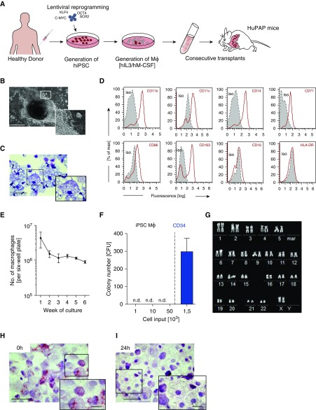
Methods: Macrophages derived from human iPSCs underwent intrapulmonary transplantation into humanized PAP mice, and engraftment, in vivo differentiation, and therapeutic efficacy of the transplanted cells were analyzed.
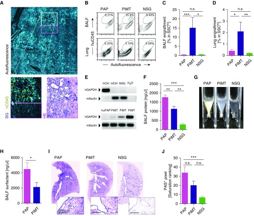
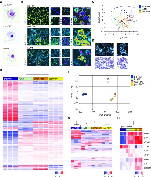
Measurements and main results: On intratracheal application, iPSC-derived macrophages engrafted in the lungs of humanized PAP mice. After 2 months, transplanted cells displayed the typical morphology, surface markers, functionality, and transcription profile of primary human alveolar macrophages. Alveolar proteinosis was significantly reduced as demonstrated by diminished protein content and surfactant protein D levels, decreased turbidity of the BAL fluid, and reduced surfactant deposition in the lungs of transplanted mice.
Conclusions: We here demonstrate for the first time that pulmonary transplantation of human iPSC-derived macrophages leads to pulmonary engraftment, their in situ differentiation to an alveolar macrophage phenotype, and a reduction of alveolar proteinosis in a humanized PAP model. To our knowledge, this finding presents the first proof-of-concept for the therapeutic potential of human iPSC-derived cells in a pulmonary disease and may have profound implications beyond the rare disease of PAP.
Keywords: cell therapy; induced pluripotent stem cell; macrophages; pulmonary alveolar proteinosis.
Figures
Comment in
-
Manmade Macrophage Offers a New Therapy for Pulmonary Alveolar Proteinosis.Am J Respir Crit Care Med. 2018 Aug 1;198(3):297-298. doi: 10.1164/rccm.201803-0478ED. Am J Respir Crit Care Med. 2018. PMID: 29669215 No abstract available.
Similar articles
-
Gene correction of human induced pluripotent stem cells repairs the cellular phenotype in pulmonary alveolar proteinosis.Am J Respir Crit Care Med. 2014 Jan 15;189(2):167-82. doi: 10.1164/rccm.201306-1012OC. Am J Respir Crit Care Med. 2014. PMID: 24279725
-
Use of induced pluripotent stem cells to recapitulate pulmonary alveolar proteinosis pathogenesis.Am J Respir Crit Care Med. 2014 Jan 15;189(2):183-93. doi: 10.1164/rccm.201306-1039OC. Am J Respir Crit Care Med. 2014. PMID: 24279752 Free PMC article.
-
Pulmonary transplantation of macrophage progenitors as effective and long-lasting therapy for hereditary pulmonary alveolar proteinosis.Sci Transl Med. 2014 Aug 20;6(250):250ra113. doi: 10.1126/scitranslmed.3009750. Sci Transl Med. 2014. PMID: 25143363
-
The influence of genetics on therapeutic developments in pulmonary alveolar proteinosis.Curr Opin Pulm Med. 2019 May;25(3):294-299. doi: 10.1097/MCP.0000000000000576. Curr Opin Pulm Med. 2019. PMID: 30865035 Review.
-
Pulmonary alveolar proteinosis: time to shift?Expert Rev Respir Med. 2015 Jun;9(3):337-49. doi: 10.1586/17476348.2015.1035259. Epub 2015 Apr 12. Expert Rev Respir Med. 2015. PMID: 25864717 Review.
Cited by
-
CSF2RB mutation-related hereditary pulmonary alveolar proteinosis: the "long and winding road" into adulthood.ERJ Open Res. 2023 Dec 18;9(6):00703-2023. doi: 10.1183/23120541.00703-2023. eCollection 2023 Nov. ERJ Open Res. 2023. PMID: 38111540 Free PMC article.
-
Studying tissue macrophages in vitro: are iPSC-derived cells the answer?Nat Rev Immunol. 2018 Nov;18(11):716-725. doi: 10.1038/s41577-018-0054-y. Nat Rev Immunol. 2018. PMID: 30140052 Review.
-
Alveolar macrophages in pulmonary alveolar proteinosis: origin, function, and therapeutic strategies.Front Immunol. 2023 Jun 14;14:1195988. doi: 10.3389/fimmu.2023.1195988. eCollection 2023. Front Immunol. 2023. PMID: 37388737 Free PMC article. Review.
-
Immunoregulatory Macrophages Modify Local Pulmonary Immunity and Ameliorate Hypoxic Pulmonary Hypertension.Arterioscler Thromb Vasc Biol. 2024 Dec;44(12):e288-e303. doi: 10.1161/ATVBAHA.124.321264. Epub 2024 Oct 10. Arterioscler Thromb Vasc Biol. 2024. PMID: 39387119
-
Unlocking the Future: Pluripotent Stem Cell-Based Lung Repair.Cells. 2024 Apr 5;13(7):635. doi: 10.3390/cells13070635. Cells. 2024. PMID: 38607074 Free PMC article. Review.
References
-
- Yu J, Vodyanik MA, Smuga-Otto K, Antosiewicz-Bourget J, Frane JL, Tian S, et al. Induced pluripotent stem cell lines derived from human somatic cells. Science. 2007;318:1917–1920. - PubMed
-
- Takahashi K, Tanabe K, Ohnuki M, Narita M, Ichisaka T, Tomoda K, et al. Induction of pluripotent stem cells from adult human fibroblasts by defined factors. Cell. 2007;131:861–872. - PubMed
-
- Takasato M, Er PX, Chiu HS, Maier B, Baillie GJ, Ferguson C, et al. Kidney organoids from human iPS cells contain multiple lineages and model human nephrogenesis. Nature. 2015;526:564–568. - PubMed
Publication types
MeSH terms
Grants and funding
LinkOut - more resources
Full Text Sources
Other Literature Sources
Research Materials
