. 2018 Aug 1;50(8):543-552.
doi: 10.1152/physiolgenomics.00118.2017. Epub 2018 Apr 13.

FAR2 is associated with kidney disease in mice and humans

Affiliations

FAR2 is associated with kidney disease in mice and humans

Grant Backer et al. Physiol Genomics. .

Abstract

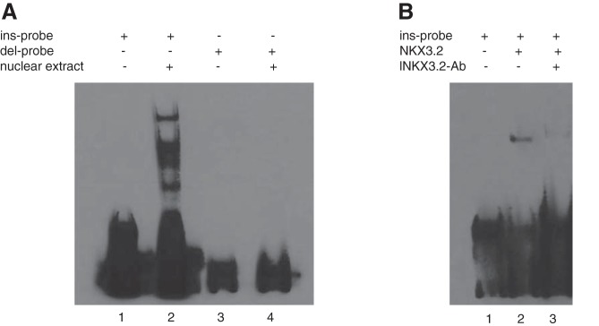
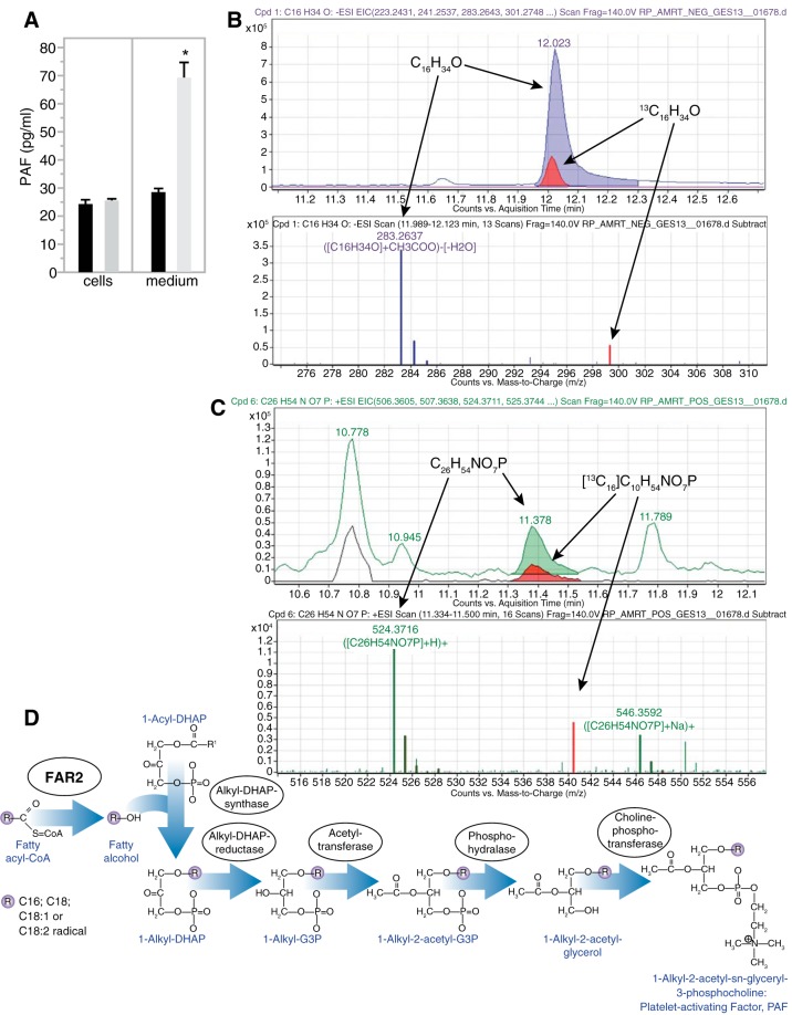
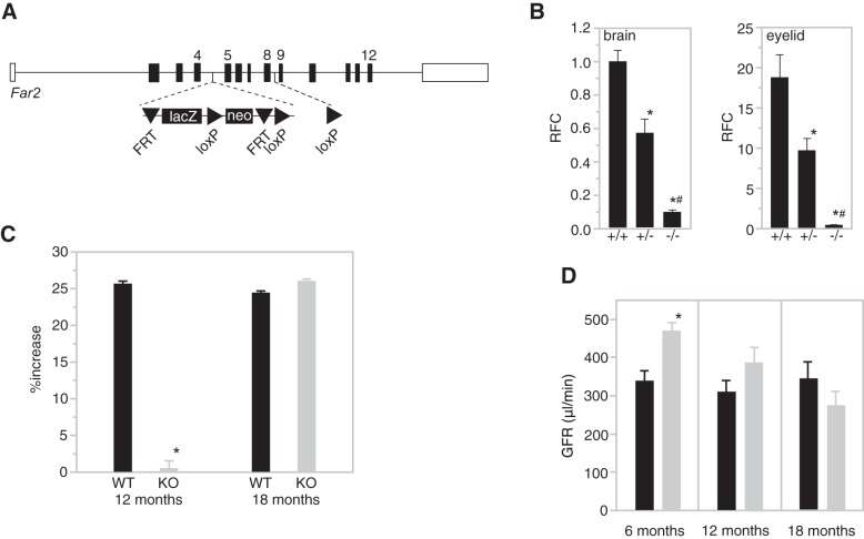
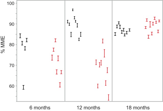
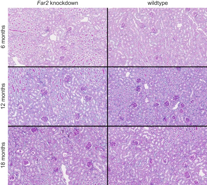
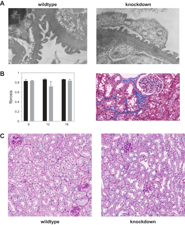
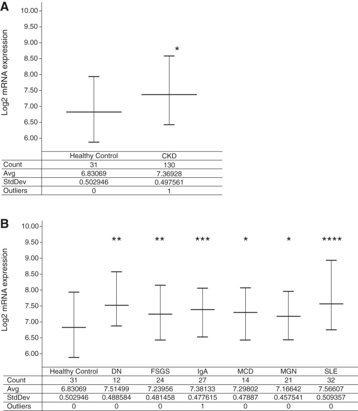
Mesangial matrix expansion is an important process in the initiation of chronic kidney disease, yet the genetic factors driving its development are unknown. Our previous studies have implicated Far2 as a candidate gene associated with differences in mesangial matrix expansion between mouse inbred strains. Consistent with the hypothesis that increased expression of Far2 leads to mesangial matrix expansion through increased production of platelet-activating factor precursors, we show that FAR2 is capable of mediating de novo platelet-activating factor synthesis in vitro and driven by the transcription factor NKX3.2. We demonstrate that knockdown of Far2 in mice delays the progression of mesangial matrix expansion with at least six months (equivalent to ~15 yr in human). Furthermore, we show that increased FAR2 expression in human patients is associated with diabetic nephropathy, lupus nephritis, and IgA nephropathy. Taken together, these results highlight FAR2's role in the development of mesangial matrix expansion and chronic kidney disease.

Keywords: aging; kidney; mesangial matrix; mouse.


Figures

Fig. 1.
Fig. 1.
The 9 bp sequence in the 5′-untranslated region (UTR) of Far2 binds NKX3-2. A: electrophoretic mobility shift assay (EMSA) performed with nuclear extract from mouse mesangial cells incubated with biotin-labeled oligonucleotide probes corresponding to the Far2 5′-UTR sequences with (ins-probe) and without (del-probe) the 9 bp sequence (lanes 2 and 4, respectively). B: EMSA performed with purified NKX3-2 protein (lane 2). A supershift assay was performed with an antibody against NKX3-2 (lane 3).
Fig. 2.
Fig. 2.
Localization of Nkx3-2 expression in the kidney by RNAScope probes. Representative images from 6, 12, and 18 mo old animals show Nkx3-2 expression (magenta) in most cells of the kidney including all cells in the glomerulus. In addition to probes for Nkx3-2, we used probes to identify the proximal tubule (Ggt, green) and the collecting duct (Aqp2, yellow). The white line indicates the outline of the glomerulus.
Fig. 3.
Fig. 3.
Far2 is an enzyme involved in de novo platelet-activating factor (PAF) production. A: PAF concentration measured in both culture media and cell lysates between Far2-overexpressing (gray, n = 3) and wild-type (black, n = 3) mesangial cells. Labeled fatty alcohol (hexadecanol) (B) and PAF (C) were detected through mass spectrometry after a mixture of 13C- and 12C-palmitate was added to Far2-overexpressing cells. D: proposed pathway highlighting the role of FAR2 in de novo synthesis of PAF.
Fig. 4.
Fig. 4.
Generation and characterization of B6N(Cg)-Far2tm2a(KOMP)Wtsi/2J. A: a simplified scheme showing the wild-type Far2 allele and knock-in allele Far2tm2a(KOMP)Wtsi. The lacZ and neomycin expression cassette was inserted between exon 4 and exon 5 and is flanked by FRT (Flippase Recognition Target) sites. Exons 5–8 are flanked by loxP sites. B: quantitative PCR for Far2 on brain and eyelid of wild-type (+/+), heterozygous (+/−), and homozygous (−/−) knockout mice (n = 3/genotype) levels were calculated as relative fold change (RFC) compared with wild type. *P < 0.01 compared with +/+; #P < 0.01 compared with +/−. C: quantification of mesangial matrix expansion (MME) in knockout mice vs. wild-type mice at 12 and 18 mo of age relative to 6 mo measurements. MME is reported as percentage of the glomerular area occupied by matrix for 50 glomeruli per mouse and 10 mice per group. P = 1.27 × 10⁻⁶. D: glomerular filtration rate (GFR) data in knockout mice (gray) vs. wild-type mice (black) at 6, 12, and 18 mo of age. n = 10 per group. *P = 0.0118.
Fig. 5.
Fig. 5.
MME data from the individual animals in the study. For each animal the %MME (average ± SE) of 50 glomeruli is shown at the 3 ages for wild-type (black) and Far2 knockdown (red) animals.
Fig. 6.
Fig. 6.
PAS staining of Far2 knockdown and wild-type kidneys at 6, 12, and 18 mo of age. These images are representative for each of the cohorts and show increased MME in wild-type animals at 12 mo compared with 6 mo (and similar compared with 18 mo), while there is no increase in MME in the knockdown animals at 12 mo compared with 6 mo of age.
Fig. 7.
Fig. 7.
Comparison between wild-type and Far2 knockdown animals for podocyte effacement (A), renal fibrosis (B), tubulo-interstitial damage, and immune cell infiltration (C). For podocyte effacement, tubulo-interstitial damage, and immune cell infiltration slides from 10 animals per group and 3 time points were scored independently by two people, and representative pictures from 12 mo old animals are shown. For renal fibrosis we quantified the fibrosis in all animals as described in materials and methods.
Fig. 8.
Fig. 8.
FAR2 expression in human patients. A: FAR2 is upregulated in the glomeruli isolated from chronic kidney disease (CKD) patients relative to healthy living donor samples. B: FAR2 is upregulated in the glomeruli isolated from diabetic nephropathy (DN), focal segmental glomerulosclerosis (FSGS), IgA nephropathy (IgA), minimal change disease (MCD), membranous glomerulonephropathy (MGN), and systemic lupus erythematosus nephritis (SLE). Significant P values are denoted by *P < 0.05, **P < 0.005, ***P < 0.0005, and ****P < 0.00001 relative to healthy living donor by a one-tailed Student’s t-test on nonlog-transformed expression data.

References

    1. Abrass CK, Adcox MJ, Raugi GJ. Aging-associated changes in renal extracellular matrix. Am J Pathol 146: 742–752, 1995. - PMC - PubMed
    1. Barrett T, Wilhite SE, Ledoux P, Evangelista C, Kim IF, Tomashevsky M, Marshall KA, Phillippy KH, Sherman PM, Holko M, Yefanov A, Lee H, Zhang N, Robertson CL, Serova N, Davis S, Soboleva A. NCBI GEO: archive for functional genomics data sets–update. Nucleic Acids Res 41, D1: D991–D995, 2013. doi:10.1093/nar/gks1193. - DOI - PMC - PubMed
    1. Cheng JB, Russell DW. Mammalian wax biosynthesis. I. Identification of two fatty acyl-Coenzyme A reductases with different substrate specificities and tissue distributions. J Biol Chem 279: 37789–37797, 2004. doi:10.1074/jbc.M406225200. - DOI - PMC - PubMed
    1. Dai M, Wang P, Boyd AD, Kostov G, Athey B, Jones EG, Bunney WE, Myers RM, Speed TP, Akil H, Watson SJ, Meng F. Evolving gene/transcript definitions significantly alter the interpretation of GeneChip data. Nucleic Acids Res 33: e175, 2005. doi:10.1093/nar/gni179. - DOI - PMC - PubMed
    1. Edgar R, Domrachev M, Lash AE. Gene Expression Omnibus: NCBI gene expression and hybridization array data repository. Nucleic Acids Res 30: 207–210, 2002. doi:10.1093/nar/30.1.207. - DOI - PMC - PubMed

Publication types

MeSH terms