. 2018 Apr 13;5(2):19.
doi: 10.3390/jcdd5020019.

Hypoxia Supports Epicardial Cell Differentiation in Vascular Smooth Muscle Cells through the Activation of the TGFβ Pathway

Affiliations

Hypoxia Supports Epicardial Cell Differentiation in Vascular Smooth Muscle Cells through the Activation of the TGFβ Pathway

Jiayi Tao et al. J Cardiovasc Dev Dis. .

Abstract

Epicardium-derived cells (EPDCs) are an important pool of multipotent cardiovascular progenitor cells. Through epithelial-to-mesenchymal-transition (EMT), EPDCs invade the subepicardium and myocardium and further differentiate into several cell types required for coronary vessel formation. We previously showed that epicardial hypoxia inducible factor (HIF) signaling mediates the invasion of vascular precursor cells critical for patterning the coronary vasculature. Here, we examine the regulatory role of hypoxia (1% oxygen) on EPDC differentiation into vascular smooth muscle cells (VSMCs).

Results: Hypoxia stimulates EMT and enhances expression of several VSMC markers in mouse epicardial cell cultures. This stimulation is specifically blocked by inhibiting transforming growth factor-beta (TGFβ) receptor I. Further analyses indicated that hypoxia increases the expression level of TGFβ-1 ligand and phosphorylation of TGFβ receptor II, suggesting an indispensable role of the TGFβ pathway in hypoxia-stimulated VSMC differentiation. We further demonstrate that the non-canonical RhoA/Rho kinase (ROCK) pathway acts as the main downstream effector of TGFβ to modulate hypoxia’s effect on VSMC differentiation.

Conclusion: Our results reveal a novel role of epicardial HIF in mediating coronary vasculogenesis by promoting their differentiation into VSMCs through noncanonical TGFβ signaling. These data elucidate that patterning of the coronary vasculature is influenced by epicardial hypoxic signals.

Keywords: RhoA/ROCK; TGFβ; coronary vasculature; epicardial cell; epithelial-to-mesenchymal-transition; hypoxia; hypoxia inducible factor; transforming growth factor beta; vascular smooth muscle cells.


Conflict of interest statement

The authors declare no conflict of interest. The funding sponsors had no role in the design of the study; in the collection, analyses, or interpretation of data; in the writing of the manuscript and in the decision to publish the results.

Figures

Figure 1
Figure 1
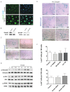
Hypoxia promoted vascular smooth muscle cell (VSMC) differentiation in epicardial cells. Immortalized mouse epicardial cells (EPDCs) were cultured under normoxic (21% O2) or hypoxic (1% O2) conditions for 24 h. In response to hypoxia, EPDCs underwent epithelial-mesenchymal transformation (EMT). Loss of cell to cell contact was evident in the zonula occludens-1 (ZO-1) labeled cells (A,B) but with no significant alterations in the expression of periostin following hypoxic treatment (C,D). Real-Time PCR (n = 3) (E) and Western blot (F) showed the induction of vascular smooth muscle cell markers, α-smooth muscle actin (SMA) and smooth muscle protein 22-alpha (SM22α) under hypoxic conditions. Hypoxia did not alter the expression of hypoxia-related genes Arnt or Hif-2α nor Flt-1 but induced the expression of a HIF transcriptional target, VefgA (n = 3) (G) HIF-1α decreased in lysates from hypoxic EPDC cultures infected with small hairpin RNA (shRNA) against HIF-1α (shHIF-1α). Scramble shRNA was used as a negative control (H). shHIF-1α repressed expression of SMA and Sm22-lacZ activity (IL) following 24 h at 1% O2. * p < 0.05; Bar: 50 μm.
Figure 2
Figure 2
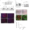
The TGFβ pathway is involved in the hypoxic induction of VSMC differentiation in EPDC cultures. ALK5/TGFβR I inhibitor SB431542 (SB) abolished the hypoxia-stimulated (1% O2, 36 h) expression of SMA (AG) and SM22α-lacZ (HN) in EPDC cultures similar to its effect on cultures treated with 250 pM TGFβ1. SM22-lacZ activity was assayed to quantify the extent of cell differentiation. Data are presented as mean fold induction of triplicate samples ± SEM compared to normoxia only conditions (n = 3) (O). Hypoxia induced TGFβ-1 and -2 and TGFβ-R3 transcript levels (n = 3) (P). ELISA data indicated that expression of TGFβ1 ligand in epicardial cells increased significantly under hypoxia by 8 and 24 h (n = 3) (Q). Hypoxia up-regulated the phosphorylation of TGFβRII (1% O2, 8h) (R). N: Normoxia; H: Hypoxia; SB: SB431542. * p < 0.05, ** p < 0.001; Bar: 50 μm.
Figure 3
Figure 3
Hypoxia mediated activation of SMADs is dispensable for differentiation of epicardial cells into VSMCs. EPDCs cultures treated with 250 pM TGFβ-1 induced nuclear-localization of Smad2, indicative of its activation (A). Western blots of EPDCs supported the knockdown of SMAD2 or SMAD4 using the appropriate siRNAs (B), which reduced the generation of SM22α-LacZ cells in TGFβ-1 treated EPDC cultures (C). Hypoxia triggered the phosphorylation of Smad2, Akt and ERK (D). Inhibition of PI3K/AKT (LY: 30 μM LY294002), MEK1/2 (UO: 10 μM UO126), or p38 MAPK (SB: 10 μM SB202190) pathways did not block hypoxia induced smooth muscle cell differentiation of EPDC cultures. Similarly, knockdown siSMAD2 or siSMAD4 also failed to block smooth muscle cell differentiation in hypoxia treated EPDC cultures (E). Quantification of SM22-LacZ+ in hypoxic EPDC cultures treated with chemical inhibitors or siRNAs showed no significant differences (n = 3) (F). Bar: 50 μm.
Figure 4
Figure 4
The RhoA/ROCK pathway acts as the main downstream effector of TGFβ to regulate VSMC differentiation in hypoxic EPDC cultures. Western blot results indicate that total RhoA levels in EPDCs are not significantly altered by hypoxia (A) but the RhoA activation (RhoA-GTP), measured by ELISA, was induced in as early as 15 min (B). phospho-SMAD2 activation by hypoxia in EPDC cultures was inhibited by selectively blocking ALK5/TGFβR I (SB; SB421542) but not with inhibitor to PI3K/AKT (LY: 30 μM LY294002) and found to increase with inhibition to ROCK (Y: 10 μM Y27632) (n = 3) * p < 0.05) (C). In contrast, inhibition of ROCK (Y) blocked SMA expression stimulated by either TGFβ (250 pM) or hypoxia, similar to blocking ALK5/TGFβR I receptor (SB: SB431542) (D). Expression of SMA in hypoxic EPDC cultures was significantly compromised by inhibiting ROCK (Y) but not PI3K/AKT (LY), even in cultures treated with exogenous TGFβ (E). Compared with Ad-GFP treated control cultures, SM22 and SMA smooth muscle cell markers were abolished in EPDCs infected with adenovirus expressing dominant negative RhoA (Ad-dnRhoA) (F). Quantification shows significantly decreased LacZ+ cell numbers in Ad-dnRhoA than Ad-GFP control treated hypoxic EPDCs cultures (n = 3) * p < 0.05 (G). N: Normoxia; H: Hypoxia; Bar: 50 μm.
Figure 5
Figure 5
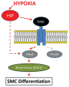
Hypoxia positively regulates epicardial cell differentiation into VSMC by the activation of the TGFβ pathway. Hypoxia promotes RhoA as the major downstream regulator of the TGFβ pathway affecting EPDC differentiation into VSMCs.

Similar articles

Cited by

References

    1. Guadix J.A., Carmona R., Munoz-Chapuli R., Perez-Pomares J.M. In vivo and in vitro analysis of the vasculogenic potential of avian proepicardial and epicardial cells. Dev. Dyn. 2006;235:1014–1026. doi: 10.1002/dvdy.20685. - DOI - PubMed
    1. Olivey H.E., Svensson E.C. Epicardial-myocardial signaling directing coronary vasculogenesis. Circ. Res. 2010;106:818–832. doi: 10.1161/CIRCRESAHA.109.209197. - DOI - PMC - PubMed
    1. Red-Horse K., Ueno H., Weissman I.L., Krasnow M.A. Coronary arteries form by developmental reprogramming of venous cells. Nature. 2010;464:549–553. doi: 10.1038/nature08873. - DOI - PMC - PubMed
    1. Tian X., Hu T., Zhang H., He L., Huang X., Liu Q., Yu W., He L., Yang Z., Zhang Z., et al. Subepicardial endothelial cells invade the embryonic ventricle wall to form coronary arteries. Cell Res. 2013;23:1075–1090. doi: 10.1038/cr.2013.83. - DOI - PMC - PubMed
    1. Tian X., Pu W.T., Zhou B. Cellular origin and developmental program of coronary angiogenesis. Circ. Res. 2015;116:515–530. doi: 10.1161/CIRCRESAHA.116.305097. - DOI - PMC - PubMed

LinkOut - more resources