Review
. 2018 Apr 13;19(4):1185.
doi: 10.3390/ijms19041185.

Excessive Extracellular ATP Desensitizes P2Y2 and P2X4 ATP Receptors Provoking Surfactant Impairment Ending in Ventilation-Induced Lung Injury

Affiliations
Review

Excessive Extracellular ATP Desensitizes P2Y2 and P2X4 ATP Receptors Provoking Surfactant Impairment Ending in Ventilation-Induced Lung Injury

Djo Hasan et al. Int J Mol Sci. .

Abstract

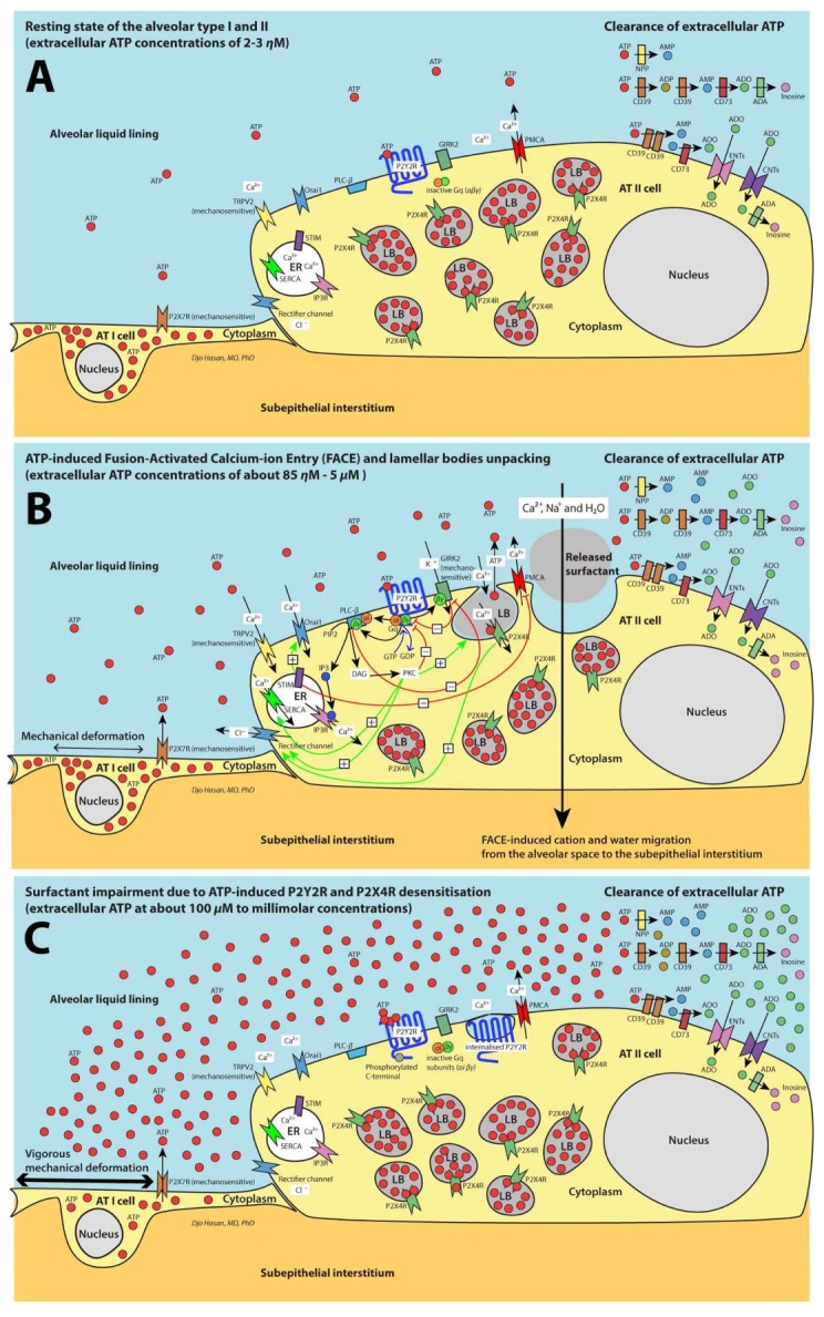
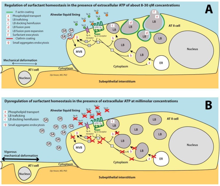
Stretching the alveolar epithelial type I (AT I) cells controls the intercellular signaling for the exocytosis of surfactant by the AT II cells through the extracellular release of adenosine triphosphate (ATP) (purinergic signaling). Extracellular ATP is cleared by extracellular ATPases, maintaining its homeostasis and enabling the lung to adapt the exocytosis of surfactant to the demand. Vigorous deformation of the AT I cells by high mechanical power ventilation causes a massive release of extracellular ATP beyond the clearance capacity of the extracellular ATPases. When extracellular ATP reaches levels >100 μM, the ATP receptors of the AT II cells become desensitized and surfactant impairment is initiated. The resulting alteration in viscoelastic properties and in alveolar opening and collapse time-constants leads to alveolar collapse and the redistribution of inspired air from the alveoli to the alveolar ducts, which become pathologically dilated. The collapsed alveoli connected to these dilated alveolar ducts are subject to a massive strain, exacerbating the ATP release. After reaching concentrations >300 μM extracellular ATP acts as a danger-associated molecular pattern, causing capillary leakage, alveolar space edema, and further deactivation of surfactant by serum proteins. Decreasing the tidal volume to 6 mL/kg or less at this stage cannot prevent further lung injury.

Keywords: P2X receptors; P2Y receptors; extracellular ATP; innate immunity; purinergic signaling; surfactant dysfunction; ventilation-induced lung injury.


Conflict of interest statement

Djo Hasan: The author reports to have received an honorarium to a lecture from Demcon-Macawi; Michaela Kollisch-Singule: The author reports to have received travel reimbursement at events sponsored by Dräger Medical; Gary F. Nieman.: The author reports to have received grants from the NIH, a grant from Dräger Medical, a grant from the CDRMP (DoD), travel and honorarium to lecture from Dräger Medical; Joshua Satalin, Philip van der Zee, Paul Blankman, Atsuko Shono, Peter Somhorst, Corstiaan den Uil, Han Meeder and Toru Kotani: The authors declare no conflicts of interest.

Figures

Figure 1
Figure 1
Schematic presentation of the regulation of surfactant exocytosis. For greater clarity, the high cytosolic adenosine triphosphate (ATP) content is omitted in the figure. (A) Resting state of the alveolar epithelial type I (AT I) and AT II cells. (B) ATP-induced fusion- activated calcium-ion entry resulting in surfactant exocytosis. (C) Excessive extracellular ATP concentrations causing the impairment of surfactant exocytosis. See text for explanation. AT I: Alveolar epithelial type I cell; AT II: Alveolar epithelial type II cell; ER: Endoplasmic reticulum; LB: Lamellar body; P2Y2R and P2X4R: ATP receptors; Gq/11: G protein-coupled receptor molecules comprising αi and βγ subunits; PLC-β: Phospholipase C beta; PIP2: Phosphatidylinositol 4,5-bisphosphate; IP3: Inositol triphosphate; IP3R: Inositol triphosphate receptor, a membrane bound glycoprotein complex functioning as a Ca2+ channel sensitive to activation by IP3; STIM1: Stromal interaction molecule 1, a calcium sensor; Orai1: Calcium release-activated calcium channel protein 1, a calcium selective ion channel; TRPV2: Transient receptor potential cation channel subfamily V member 2, a non-selective cation channel; Kir3: K+ selective inwardly rectifying channel 3 or GIRK2: G protein-coupled inwardly-rectifying K+ channel 2; DAG: diacylglycerol; PKC: protein kinase C; CD39: Nucleoside triphosphate diphosphohydrolase 1 (NTPD1); NPP: nucleotide pyrophosphatase/phosphodiesterase; CD73: 5′-nucleotidase (5′-NT); ADA: adenosine deaminase; ENTs: Equilibrative nucleoside transporters 1 and 2; CNTs: Concentrative nucleoside transporters 1 and 2; FACE: fusion-activated Ca2+ entry; SERCA: sarcoplasmic/endoplasmic reticulum Ca2+ ATPase channel; PMCA: Plasma membrane Ca2+ ATPase channel. Figures extensively adapted from Hasan, et al. (2017) [10] (open access) with permission.
Figure 2
Figure 2
Schematic presentation of the surfactant homeostasis of the alveolar epithelial cells. (A) A perivesicular F-actin coating is formed around the fused LBs after the initial LB fusion pore has developed. Several types of fusion pore development are described [57]: (1) 80% of the F-actin-coated fused LBs release surfactant and the LB membrane becomes part of the plasma membrane (kiss-coat-and-release) followed by the disappearance of the F-actin coat; (2) 10% of the F-actin-coated fused LBs discontinued the fusion process and returned inside the cell (kiss-coat-and-run); (3) In the remaining F-actin-coated LBs the fusion process was arrested for a certain time (<20 min) (kiss-coat-and-wait) [57]. The endocytosis of SAs occurs through a clathrin-dependent pathway [59] by the activation of several types of SP-A receptors [59,60,61,62,63] and a SP-D receptor [64]. The SP-D receptor is a GPR116, also known as Ig-Hepta that are highly expressed in the lung [64]. Besides SAs uptake, this process also inhibits the surfactant exocytosis and contributes to the control of extracellular surfactant homeostasis [59,64]. (B) The activation of the pro-inflammatory response of the innate immune system through the activation of the P2X7Rs by extracellular ATP at >300 μM concentrations (ATP molecules at these concentrations act as DAMPs) leads to the recruitment and activation of neutrophils. The recruited and activated neutrophils cause the degradation of SP-D and SP-A leading to a deficiency of SP-D and SP-A [65,66] preventing the clathrin-dependent recycling of the majority of SAs and aborting the above-mentioned inhibition of the trafficking, semi-fusion and fusion of the LBs with the cell membrane. AT I: Alveolar epithelial type I cell; AT II: Alveolar epithelial type II cell; ER: Endoplasmic reticulum; LB: Lamellar body; MVB: multivesicular body; SP-A: Surfactant protein A; SP-D: Surfactant protein D; SAs: Surfactant small aggregates; LAs: Surfactant large aggregates; GPR116: G protein-coupled receptor 116; DAMPs: danger-associated molecular patterns.
Figure 3
Figure 3
Graphic presentation of the time course of alveolar collapse during the expiration by releasing an airway pressure of 25 cm H2O to zero as depicted by in-vivo microscopy in rats with surfactant-deactivated lung. The Y-axis represents the alveolar surface areas in pixels and the X-axis is the time. There is a time lag of 0.17 s before alveoli start to collapse after the initiation of the expiratory phase. Furthermore, it takes 0.25 s before the alveoli are fully collapsed. Figure from Satalin, et al. (2016) [103], presented at ‘The Open Forum Sessions’ during the AARC Congress 2016.
Figure 4
Figure 4
The effect of the ventilator settings on the alveolar mechanics. The left graphics are schematic presentations of the ventilator pressure-time curves belonging to the photomicrographs of the lung presented on the right figure. The lung was fixed at the end-inspiratory pressure (left column of the photomicrographs) and at the end-expiratory pressure (right column of the photomicrographs). The conducting airspaces including the alveolar ducts are colored green, the alveolar spaces are magenta and the alveolar walls are lilac. In APRV75% and APRV10% termination of the expiration is set at an EEF/PEF ratio of 75% and 10%, respectively. In the healthy lung using tissue microscopy after fixing the lung at peak-inspiration and at end-expiration, Kollisch-Singule, et al. (2014) demonstrated that the distribution of tidal volume between the alveoli and the alveolar ducts shows little change during inspiration and expiration (‘control’) [100]. After surfactant deactivation, there is a redistribution of air at the end of expiration from the alveoli towards the alveolar ducts (‘expiration’ and ‘PEEP 5’). During inspiration, the redistribution towards the alveolar ducts markedly increases causing a tremendous deformation of the alveoli adjacent to these alveolar ducts (‘inspiration’ and ‘PEEP 5’). This results in an increased microstrain (defined as the change in length of the alveolar ducts between inspiration and expiration normalized by their original length). Increasing the PEEP level to 16 cm H2O decreases the microstrain but not the redistribution of air towards the alveolar ducts (‘Inspiration’, ‘expiration’ and ‘PEEP 16’). The application of APRV10% with a Phigh of 40 cm H2O and expiratory time of 0.22–0.26 s increases the redistribution of air towards the alveolar ducts and the microstrain dramatically (‘Inspiration’, ‘expiration’ and ‘APRV10%’). By applying APRV75% with a Phigh of 40 cm H2O with a shorter expiration time 0f 0.04 to 0.08 s the redistribution of air towards the alveolar ducts and the microstrain much improve but are still not completely restored (‘Inspiration’, ‘expiration’ and ‘APRV75%’) [100]. Thus: in surfactant deactivated lung, a short expiratory time stabilizes the alveoli and a long expiratory time allows alveolar collapse to occur. By setting the timing of the termination of the expiration relative the PEF, the actual expiration time will change proportional to the time-constant of the alveoli. For instance, in slowly deflating alveoli a longer time is required to reach an EEF/PEF ratio of 75% than in fast deflating alveoli. Consequently, the expiration time in a lung with a high compliance is longer than in a lung with a low compliance. Therefore, this mode is now referred to as the ‘time-controlled adaptive ventilation’ (TCAV). APRV: airway pressure release ventilation; EEF: end-expiratory flow; PEF: peak-expiratory flow; Phigh: inspiratory pressure; PEEP: positive end expiratory pressure; Vt: tidal volume; RR: respiratory rate; Exp: Expiratory. Photomicrographs figure from Kollisch-Singule, et al. (2014) [100] with permission.

References

    1. Lohmann K. Über die Pyrophosphatfraktion im Muskel. Naturwissenschaften. 1929;17:624–625. doi: 10.1007/BF01506215. - DOI
    1. Langen P., Hucho F. Karl Lohmann and the discovery of ATP. Angew. Chem. Int. Ed. Engl. 2008;47:1824–1827. doi: 10.1002/anie.200702929. - DOI - PubMed
    1. Williams R.J. The history and the hypotheses concerning ATP-formation by energised protons. FEBS Lett. 1978;85:9–19. doi: 10.1016/0014-5793(78)81238-1. - DOI - PubMed
    1. Wikstrom M.K., Saari H.T. Conformational changes in cytochrome aa3 and ATP synthetase of the mitochondrial membrane and their role in mitochondrial energy transduction. Mol. Cell. Biochem. 1976;11:17–33. doi: 10.1007/BF01792831. - DOI - PubMed
    1. Feldberg W., Hebb C. The stimulating action of phosphate compounds on the perfused superior cervical ganglion of the cat. J. Physiol. 1948;107:210–221. doi: 10.1113/jphysiol.1948.sp004264. - DOI - PMC - PubMed

MeSH terms

Substances

LinkOut - more resources