Review
. 2018 Apr 13;19(4):1188.
doi: 10.3390/ijms19041188.

Sympathetic Nerve Hyperactivity in the Spleen: Causal for Nonpathogenic-Driven Chronic Immune-Mediated Inflammatory Diseases (IMIDs)?

Affiliations
Review

Sympathetic Nerve Hyperactivity in the Spleen: Causal for Nonpathogenic-Driven Chronic Immune-Mediated Inflammatory Diseases (IMIDs)?

Denise L Bellinger et al. Int J Mol Sci. .

Abstract

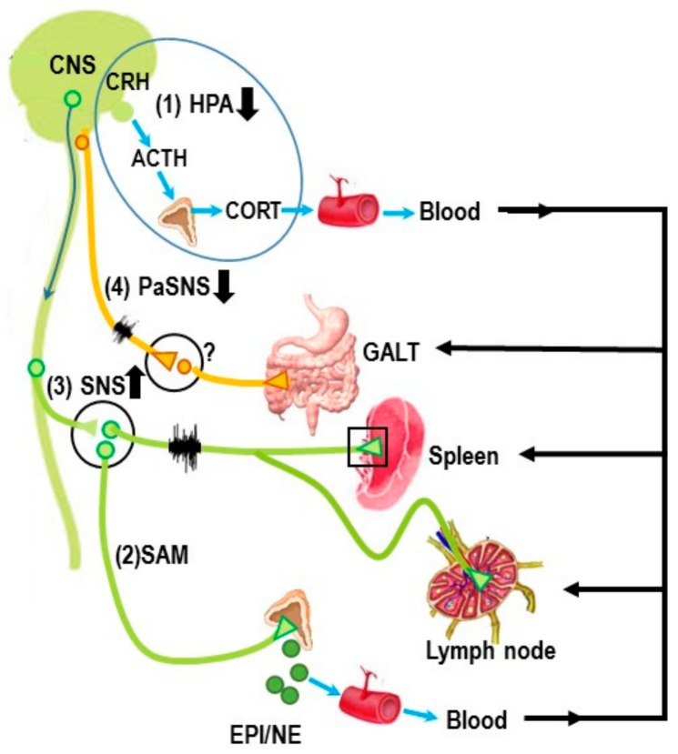
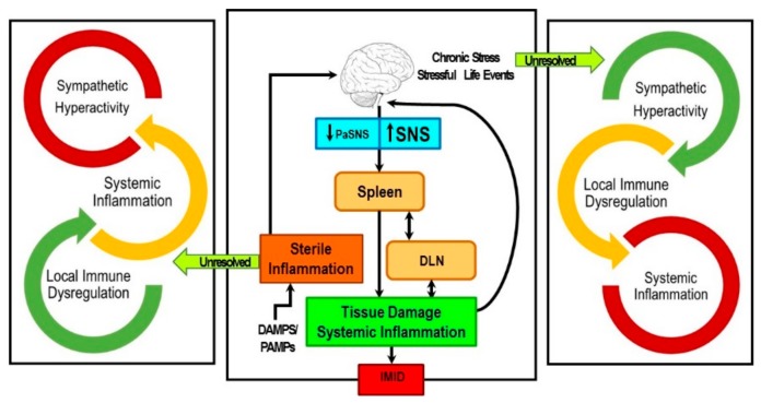
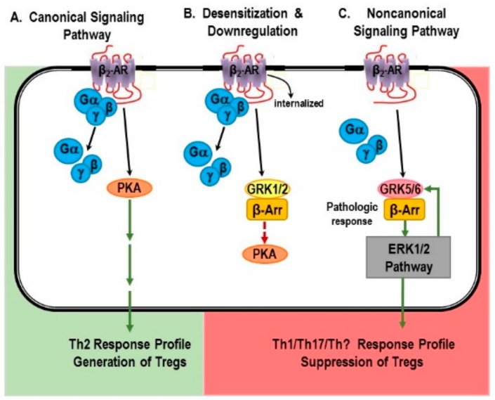
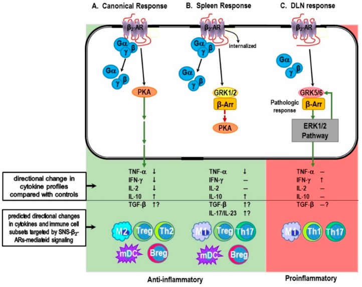
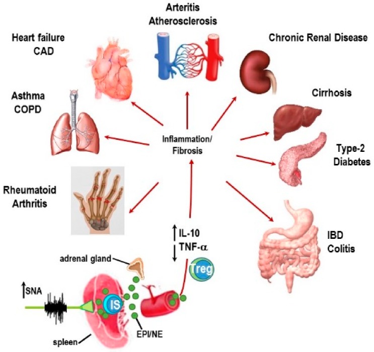
Immune-Mediated Inflammatory Diseases (IMIDs) is a descriptive term coined for an eclectic group of diseases or conditions that share common inflammatory pathways, and for which there is no definitive etiology. IMIDs affect the elderly most severely, with many older individuals having two or more IMIDs. These diseases include, but are not limited to, type-1 diabetes, obesity, hypertension, chronic pulmonary disease, coronary heart disease, inflammatory bowel disease, and autoimmunity, such as rheumatoid arthritis (RA), Sjőgren's syndrome, systemic lupus erythematosus, psoriasis, psoriatic arthritis, and multiple sclerosis. These diseases are ostensibly unrelated mechanistically, but increase in frequency with age and share chronic systemic inflammation, implicating major roles for the spleen. Chronic systemic and regional inflammation underlies the disease manifestations of IMIDs. Regional inflammation and immune dysfunction promotes targeted end organ tissue damage, whereas systemic inflammation increases morbidity and mortality by affecting multiple organ systems. Chronic inflammation and skewed dysregulated cell-mediated immune responses drive many of these age-related medical disorders. IMIDs are commonly autoimmune-mediated or suspected to be autoimmune diseases. Another shared feature is dysregulation of the autonomic nervous system and hypothalamic pituitary adrenal (HPA) axis. Here, we focus on dysautonomia. In many IMIDs, dysautonomia manifests as an imbalance in activity/reactivity of the sympathetic and parasympathetic divisions of the autonomic nervous system (ANS). These major autonomic pathways are essential for allostasis of the immune system, and regulating inflammatory processes and innate and adaptive immunity. Pathology in ANS is a hallmark and causal feature of all IMIDs. Chronic systemic inflammation comorbid with stress pathway dysregulation implicate neural-immune cross-talk in the etiology and pathophysiology of IMIDs. Using a rodent model of inflammatory arthritis as an IMID model, we report disease-specific maladaptive changes in β₂-adrenergic receptor (AR) signaling from protein kinase A (PKA) to mitogen activated protein kinase (MAPK) pathways in the spleen. Beta₂-AR signal "shutdown" in the spleen and switching from PKA to G-coupled protein receptor kinase (GRK) pathways in lymph node cells drives inflammation and disease advancement. Based on these findings and the existing literature in other IMIDs, we present and discuss relevant literature that support the hypothesis that unresolvable immune stimulation from chronic inflammation leads to a maladaptive disease-inducing and perpetuating sympathetic response in an attempt to maintain allostasis. Since the role of sympathetic dysfunction in IMIDs is best studied in RA and rodent models of RA, this IMID is the primary one used to evaluate data relevant to our hypothesis. Here, we review the relevant literature and discuss sympathetic dysfunction as a significant contributor to the pathophysiology of IMIDs, and then discuss a novel target for treatment. Based on our findings in inflammatory arthritis and our understanding of common inflammatory process that are used by the immune system across all IMIDs, novel strategies to restore SNS homeostasis are expected to provide safe, cost-effective approaches to treat IMIDs, lower comorbidities, and increase longevity.

Keywords: G protein receptor kinase; adrenergic receptor signaling; immune-mediated inflammatory diseases; inflammatory reflex; mitogen-activated protein kinase; neural-immune; protein kinase A; rheumatoid arthritis; sympathetic nervous system.


Conflict of interest statement

The authors declare no conflict of interest.

Figures

Figure 1
Figure 1
The major neuroendocrine and autonomic pathways that regulate secondary immune organs are illustrated. They include two neuroendocrine pathways, (1) the hypothalamic–pituitary–adrenal axis (HPA) and (2) the sympatho-adrenal medullary (SAM) axis, and two “hardwired” autonomic circuits, (3) the sympathetic nervous system (SNS) and (4) the parasympathetic nervous system (PaSNS). (1) The HPA axis is a feedback system whereby corticotropin-releasing hormone (CRH) is released from neurons in the hypothalamus that project their axons into the anterior pituitary. The release of CRH stimulates the secretion of adrenocorticotropic hormone (ACTH) into the circulation. ACTH stimulates the release of corticosterone (CORT) into the bloodstream. Circulating CORT has systemic effects including effects on secondary immune organs or tissues (Spleen, lymph nodes and gut-associated lymphoid tissue (GALT) are shown in the illustration). (2) In the SAM axis, preganglionic sympathetic neurons secrete acetylcholine in the adrenal medulla, which stimulates the release of catecholamines, predominantly epinephrine (EPI) and, to a much lesser extent, norepinephrine (NE), into the circulation, such as the HPA axis. Circulating NE/EPI can potentiate the actions of sympathetic nerves acting at adrenergic receptors in immune organs. (3) The SNS circuit to the spleen and LNs is a two-neuron chain (green neurons), with preganglionic cholinergic sympathetic neurons innervating postganglionic neurons whose nerve terminals end in the spleen and lymph nodes. The major neurotransmitter of postganglionic neurons is norepinephrine (NE). In secondary lymphoid organs, such as LNs and spleen, NE binds to adrenergic receptors expressed on immune cells, vasculature and connective tissue cells. Ligand binding to adrenergic receptors regulates the cellular responses of the immune cells that express these receptors. The predominant adrenergic receptor subtype expressed on spleen cells is the β2-adrenergic receptor. (4) The PaSNS is also a two-neuron chain (yellow neurons) of efferent nerves that supply the viscera, in particular the gut. Preganglionic neurons reside in the brainstem (medulla) and exit the central nervous system (CNS) as cranial nerves that end upon postganglionic neurons, most often embedded in the visceral organs they supply. Depicted here is the parasympathetic supply to gut-associated lymphoid tissue (GALT). There is no definitive evidence that the PaSNS innervates the spleen, but the vagus nerve clearly can influence immune responses in the spleen—most likely via the migration of vagally influenced immune cells in the gut that migrate to the spleen. The up or down arrows shown next to these regulatory pathways indicate the most common direction of change in the activity of that pathway in most of the prevalent immune-mediated inflammatory diseases, including rheumatoid arthritis.
Figure 2
Figure 2
Chronic stress and/or stressful adverse life events chronically elevate sympathetic nervous system (SNS) activity and reactivity that can induce low grade local and systemic inflammation. Conversely, activation of the immune system by sterile inflammagens (danger-associated molecular patterns (DAMPs) and/or pathogen-associated molecular patterns (PAMPs)) further activates the SNS. Unresolved local and/or systemic inflammation increases SNS activity. Hyper-SNS activity (hyper-SNA) can influence/skew immune function toward a functional phenotype that can trigger or worsen immune-mediated inflammatory diseases (IMIDs), which promote visceral organ and sympathetic nerve damage. Typically, the parasympathetic nervous system (PaSNS) is suppressed. Hyper-SNA in the spleen and unresolved sterile inflammation co-conspire to precipitate and/or perpetuate an IMID. Lymph nodes drain the site of tissue damage (DLN) and influence the spleen via a lymph that drains into the spleen, and activated immune cells in the spleen migrate to the DLNs.
Figure 3
Figure 3
Normal β2-adrenergic receptor (β2-AR) signal transduction via G-coupled proteins (canonical), β2-AR desensitization and downregulation, and alternative signal transduction mediated by β-arrestin (β-Arr) are shown in (AC), respectively. (A) The Canonical pathway: Ligand binding to the β2-AR causes dissociation of G-protein to Gα and Gβγ and activation of protein kinase A (PKA). Traditionally, activation of this signaling pathway promotes a Th2-type immune response and an increase in Treg cell function that promotes immune response resolution and suppression of inflammation. (B) Desensitization and Downregulation: Chronically high SNA in the spleen desensitizes and down-regulates β2-AR via G protein-coupled receptor kinase (GRK)1/2/β-arrestin (β-Arr) signal transduction that inhibits the p38MAPK pathway, ERK1/2, which prevents PKA activity. (C) The activation of the non-canonical β2-AR signaling pathway is mediated by a non-G protein mechanism (G protein is uncoupled to the receptor). Instead, GRK5/β-arrestin (β-Arr) signal transduction activates the ERK1/2 signaling pathways. The consequences for these two signaling pathways for immune responses in the spleen are indicated below. Signal transduction by the PKA pathway promotes a Th2-type immune response and the generation of Treg cells. In contrast, β2-AR signaling via the ERK1/2 pathway drives Th1 and/or Th17 immune responses depending on the immune stimulus, and suppression of Treg cell function.
Figure 4
Figure 4
(A) Canonical β2-adrenergic receptor (β2-AR) signaling response, the anti-inflammatory cytokine profile mediated by protein kinase A (PKA), and the major anti-inflammatory cellular mediators of the resulting anti-inflammatory immune response. (B) The physiological G protein-coupled receptor kinase (GRK)1/2-mediated β2-AR desensitization/downregulation in spleen cells from rats with inflammatory arthritis, the reported directional changes in cytokine profiles compared with controls, and the predicted cellular mediators of the resulting immune response. (C) The pathological GRK5/6-mediated activation of the ERK1/2 pathway in immune cells from lymph node cells that drain the affected joints (DLN cells) from rats with inflammatory arthritis, the reported directional changes in cytokine profiles compared with controls, and the predicted cellular mediators of the resulting immune response. Predicted immune cell mediators are based on well-documented functions and cytokine profiles secreted by specific populations of immune cells, particularly the secretion of the anti-inflammatory cytokines, interleukin (IL)-10 and tumor growth factor-β (TGF-β) by regulatory B and T cells (Breg and Treg, respectively), IL-10 by myeloid dendritic cells (mDC), tumor necrosis factor (TNF)-α by macrophages, IL-2 by Th2 cells, and interferon (IFN)-γ by T-helper (Th)1 and Th17/Th1 cells. Predicted M1 and M2 macrophages are based on their ability to drive a Th1/Th17 or Th2-type immune responses. SNS, sympathetic nervous system.
Figure 5
Figure 5
One mechanism is proposed whereby high sympathetic nerve activity (SNA) in the spleen may dampen systemic inflammation (red arrows) in immune-mediated inflammatory diseases (IMIDs). In the spleen, a β2-AR-mediated increase of interleukin-10 (IL-10, upward arrow) secretion and inhibition of tumor necrosis factor (TNF-α, downward arrow) by splenic immunocytes into the systemic circulation exert anti-inflammatory actions in tissues and organs (indicated by red arrows) that are targeted by IMIDs, including inflammatory bowel diseases (IBD) such as colitis, type-2 diabetes, cirrhosis, chronic renal disease, arteritis, atherosclerosis, chronic heart failure, coronary artery disease (CAD), asthma, chronic obstructive pulmonary disease (COPD), and rheumatoid arthritis.

References

    1. Rahman P., Inman R., El-Gabalawy H., Krause D.O. Pathophysiology and pathogenesis of immune-mediated inflammatory diseases: Commonalities and differences. J. Rheumatol. Suppl. 2010;85:11–26. doi: 10.3899/jrheum.091462. - DOI - PubMed
    1. Bellinger D.L., Lorton D. Autonomic regulation of cellular immune function. Auton. Neurosci. 2014;182:15–41. doi: 10.1016/j.autneu.2014.01.006. - DOI - PubMed
    1. Downing J.E., Miyan J.A. Neural immunoregulation: Emerging roles for nerves in immune homeostasis and disease. Immunol. Today. 2000;21:281–289. doi: 10.1016/S0167-5699(00)01635-2. - DOI - PubMed
    1. McEwen B.S. Sex, stress and the hippocampus: Allostasis, allostatic load and the aging process. Neurobiol. Aging. 2002;23:921–939. doi: 10.1016/S0197-4580(02)00027-1. - DOI - PubMed
    1. Elenkov I.J., Iezzoni D.G., Daly A., Harris A.G., Chrousos G.P. Cytokine dysregulation, inflammation and well-being. Neuroimmunomodulation. 2005;12:255–269. doi: 10.1159/000087104. - DOI - PubMed

LinkOut - more resources