. 2018 Apr 13;13(4):e0195845.
doi: 10.1371/journal.pone.0195845. eCollection 2018.

RhoGAPp190: A potential player in tbph-mediated neurodegeneration in Drosophila

Affiliations

RhoGAPp190: A potential player in tbph-mediated neurodegeneration in Drosophila

Simona Langellotti et al. PLoS One. .

Abstract

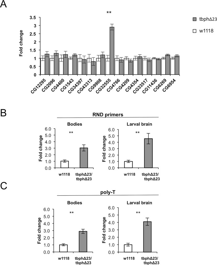
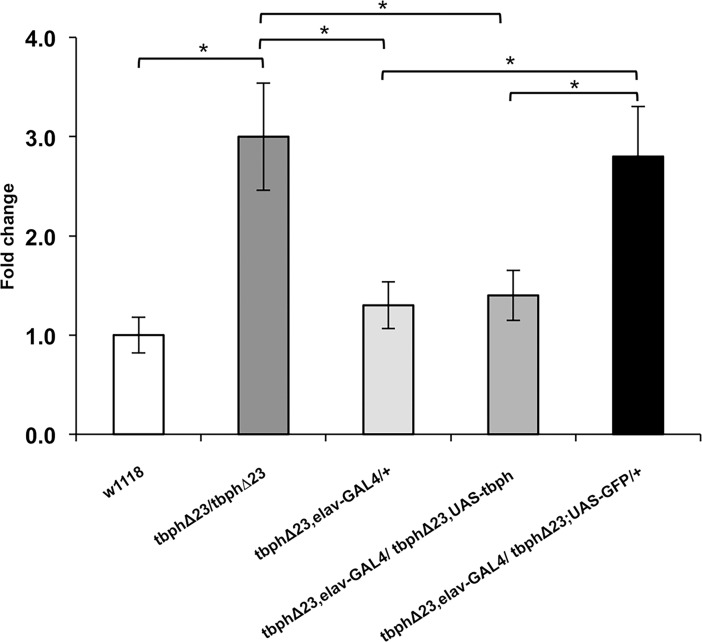
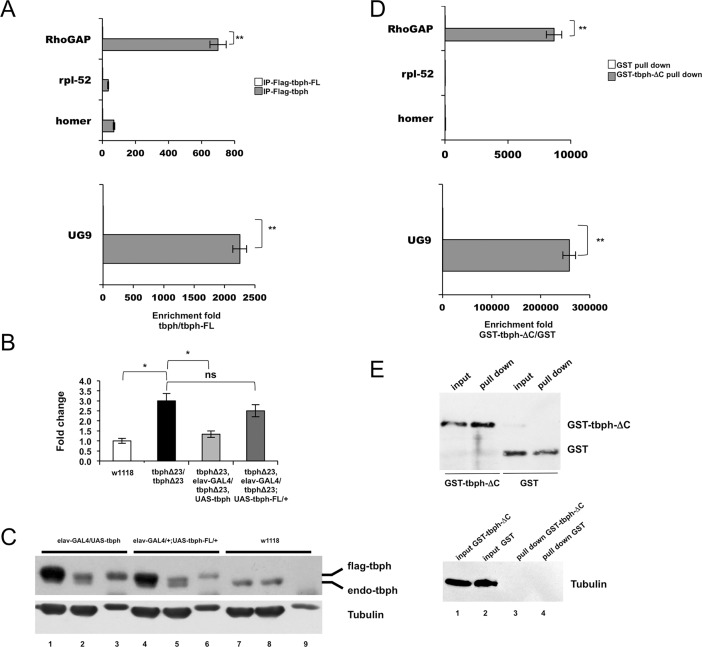
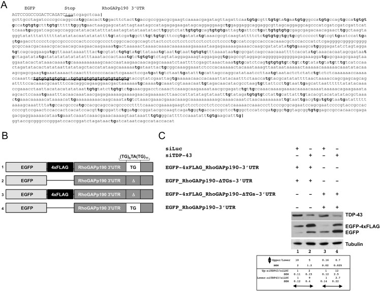
TDP-43 is an ubiquitous and highly conserved ribonucleoprotein involved in several cellular processes including pre-mRNA splicing, transcription, mRNA stability and transport. Notwithstanding the evidence of TDP-43 involvement in the pathogenesis of different neurodegenerative disorders (i.e. ALS and FTLD), the underlying mechanisms are still unclear. Given the high degree of functional similarity between the human and fly orthologs of TDP-43, Drosophila melanogaster is a simple and useful model to study the pathophysiological role of this protein in vivo. It has been demonstrated that the depletion of the TDP-43 fly ortholog (tbph) induces deficient locomotive behaviors and reduces life span and anatomical defects at the neuromuscular junction. In this study, using the known binding specificity of TDP-43/tbph for (UG) repeated sequences, we performed a bioinformatic screening for fly genes with at least 6 (TG) repeats in a row within the 3'-UTR regions in order to identify the genes that might be regulated by this factor. Among these genes, we were able to identify RhoGAPp190 as a potential target of the tbph-mediated neurodegeneration. RhoGAPp190 is a negative regulator of Drosophila RhoA, a GTPase protein implicated in the fine modulation of critical cellular processes including axon branch stability and motor axon defasciculation at muscle level and cognitive processes. We were able to demonstrate that the RhoGAPp190 expression is upregulated in a tbph-null fly model, providing evidence that this deregulation is associated to tbph silencing. Our results introduce RhoGAPp190 as a novel potential mediator in the complex scenario of events resulting from in vivo tbph loss-of-function.


Conflict of interest statement

Competing Interests: The authors have declared that no competing interests exist.

Figures

Fig 1
Fig 1. 3'UTR-TG-gene expression comparison in tbph-null and control flies.
A) Real Time PCR on RNA from fly heads samples: only the RhoGAPp190 mRNA (gene ID: CG32555) is significantly upregulated in the heads of tbph-null flies (tbphΔ23 genotype, gray bars) in comparison to controls (w1118 genotype, white bars). N = 3, **p<0.01 (t-test), error bars indicate SEM. The transcript’s expression difference was not statistically significant for the other tested genes. B) Real Time PCR carried out by using cDNA synthesized with Random primers (RND primers) or C) Real Time PCR carried out by using cDNA synthesized with poly-T primers (poly-T). Bodies samples: qPCR results show that RhoGAPp190 mRNA levels are upregulated also in the body of tbph-null flies. Larval brain: qPCR results show that RhoGAPp190 mRNA levels are upregulated also in the brain of tbph-null larvae (tbphΔ23), always in comparison to the w1118 line (w1118) used as a control. W1118 genotype, white bars; tbphΔ23 genotype, gray bars. Y-axis shows the relative fold change (tbphΔ23/w1118). N = 3, **p<0.01 (t-test), error bars indicate SEM.
Fig 2
Fig 2. Correlation of RhoGAPp190 mRNA expression with tbph expression.
Real Time PCR performed on fly heads: the mRNA levels are shown as relative expression vs w1118 genotype (= 1). In vivo tbph UAS-GAL4-driven overexpression in neurons (tbphΔ23, elav-GAL4/tbphΔ23,UAS-tbph) rescues RhoGAPp190 mRNA to the levels observed in control flies (tbphΔ23,elav-GAL4/+). GFP expression in tbph-null flies (tbphΔ23,elav-GAL4/tbphΔ23;UAS-GFP/+) was used as a control experiment. The expression in w1118 and tbph-null flies (tbphΔ23/tbphΔ23) is also shown. N = 3, *p<0.05 (t-test), error bars indicate SEM.
Fig 3
Fig 3. Genetic interaction between tbph and RhoGAPp190.
A) The climbing ability of tbph-hypomorphic allele flies is partially rescued in flies silenced for RhoGAPp190 (tbhypo/6429: tbphΔ23,elav-GAL4/UAS-RhoGAPp190-RNAi;UAS-Dcr2,UAS-tbph-RNAi/+) and (tbhypo/6430: tbphΔ23,elav-GAL4/+;UAS-Dcr2,UAS-tbph-RNAi/UAS-RhoGAPp190-RNAi) flies with respect to the animals expressing an unrelated protein (tbhypo/GFP: tbphΔ23,elav-GAL4/+;UAS-Dcr2,UAS-tbph-RNAi/UAS-GFP). The assay was performed on 4 day-old adult animals. B) Effect of single RNAi overexpression in a wild type background (the behavior of w1118 and OregonR flies was overlapping). The observed variations were not statistically significant (ns). w1118 = white 1118. OR = OregonR. (GFP = elav-GAL4/+;UAS-GFP/UAS-Dcr-2). (6429 = elav-GAL4/RhoGAPp190 RNAi; UAS-Dcr-2/+). (6430 = elav-GAL4/+; RhoGAPp190 RNAi/Dcr-2). 100 flies for each genotype were tested. *p<0.05 (t-test), error bars indicate SEM.
Fig 4
Fig 4. Probing tbph/RhoGAPp190 mRNA interaction.
A) Co-immunoprecipitation. The RhoGAPp190 mRNA was immunoprecipitated with Flag-tagged tbph or with Flag-tagged tbph-FL from transgenic fly head extracts. The UG9 RNA was used as positive control. Homer and rpl-52 transcripts were used negative controls (not statistically significant). The enrichment-fold, quantified by Real Time PCR, is referred to the mutant tbph-FL (ratio tbph/tbph-FL). N = 3, **p<0.01 (t-test), error bars indicate SEM. B) Real Time PCR performed on fly heads to test the effects of tbph-FL overexpression on RhoGAPp190 mRNA expression: in vivo tbph overexpression in neurons (tbphΔ23,elav-GAL4/ tbphΔ23,UAS-tbph), by using the UAS-GAL4 system rescues the RhoGAPp190 mRNA levels to those observed in w1118 controls. The tbph-FL overexpression in tbph-null flies (tbphΔ23,elav-GAL4/ tbphΔ23;UAS-tbph-FL/+) did not significantly affect RhoGAPp190 expression. N = 3, **p<0.01 (t-test), error bars indicate SEM. C) Co-immunoprecipitation efficiency tested by Western blot. Head extracts from elav-GAL4/UAS-tbph, elav-GAL4/+;UAS-tbph-FL/+ and w1118 flies were probed with an anti-tbph antibody. Lanes 1, 4 and 7 = input; Lanes 2, 5 and 8 = supernatant not bound after incubation with beads/antibody; Lanes 3, 6 and 9 = co-IP samples. With the anti-tbph serum, both endogenous (endo-tbph) and flag-tagged tbph (flag-tbph) are visualized. D) GST pull-down. The RhoGAPp190 mRNA was precipitated with recombinant GST-tagged tbph or GST proteins. The UG9 RNA was used as positive control. Homer and rpl-52 transcripts were used as negative controls (not statistically significant). The enrichment-fold, quantified by Real Time PCR, is referred to the GST protein (ratio GST-tbph/GST). N = 3, **p<0.01 (t-test), error bars indicate SEM. E) GST pull down efficiency tested by Western blot. Head extracts from w1118 flies were probed with an anti-GST antibody before (input) and after pull down (pull down) with GST proteins. Western blot anti-Tubulin (Tubulin) was used as a loading control for input cell extracts.
Fig 5
Fig 5. TDP-43 is implicated in the regulation of RhoGAPp190 expression.
A) Sequence of the 3’UTR of RhoGAPp190 cloned in the pEGFP-C2 vector. All TG dinucleotide repeats within the 3'UTR are boldface. The TG-repeat rich sequence located 1470nts downstream of the stop codon is underlined. B) Schematic representation of the EGFP- RhoGAPp190-3’UTR constructs. All relevant elements within the construct are shown. C) Effect of TDP-43 depletion on EGFP-RhoGAPp190-3’UTR constructs. Equal amounts of the indicated EGFP-RhoGAPp190 constructs were co-transfected in Hek293 cells. The effects of TDP-43 silencing (siTDP-43) on EGFP expression were monitored by anti-GFP immunoblotting. Anti-luciferase siRNA (siLUC) was used as the control. Western blot anti-tubulin (Tubulin) served as the protein loading control. One representative blot is shown of three independent experiments. Quantification of protein levels are shown as relative ratio of upper vs lower band in each condition or of siTDP vs siLUC treatment. Values are expressed in mean (of three independent experiments) ± SEM. P<0.05.

References

    1. Ratti A, Buratti E. Physiological functions and pathobiology of TDP-43 and FUS/TLS proteins. J Neurochem. 2016;138 Suppl 1:95–111. doi: 10.1111/jnc.13625 . - DOI - PubMed
    1. Arai T, Hasegawa M, Akiyama H, Ikeda K, Nonaka T, Mori H, et al. TDP-43 is a component of ubiquitin-positive tau-negative inclusions in frontotemporal lobar degeneration and amyotrophic lateral sclerosis. Biochem Biophys Res Commun. 2006;351(3):602–11. Epub 2006/11/07. S0006-291X(06)02318-7 [pii] doi: 10.1016/j.bbrc.2006.10.093 . - DOI - PubMed
    1. Neumann M, Sampathu DM, Kwong LK, Truax AC, Micsenyi MC, Chou TT, et al. Ubiquitinated TDP-43 in frontotemporal lobar degeneration and amyotrophic lateral sclerosis. Science. 2006;314(5796):130–3. doi: 10.1126/science.1134108 . - DOI - PubMed
    1. Ling SC, Polymenidou M, Cleveland DW. Converging mechanisms in ALS and FTD: disrupted RNA and protein homeostasis. Neuron. 2013;79(3):416–38. doi: 10.1016/j.neuron.2013.07.033 ; PubMed Central PMCID: PMC4411085. - DOI - PMC - PubMed
    1. Van Langenhove T, van der Zee J, Van Broeckhoven C. The molecular basis of the frontotemporal lobar degeneration-amyotrophic lateral sclerosis spectrum. Annals of medicine. 2012;44(8):817–28. doi: 10.3109/07853890.2012.665471 ; PubMed Central PMCID: PMC3529157. - DOI - PMC - PubMed

Publication types

MeSH terms

LinkOut - more resources