

Potential Impact of 68Ga-PSMA-11 PET/CT on the Planning of Definitive Radiation Therapy for Prostate Cancer
- PMID: 29653978
- PMCID: PMC6225538
- DOI: 10.2967/jnumed.118.209387
Potential Impact of 68Ga-PSMA-11 PET/CT on the Planning of Definitive Radiation Therapy for Prostate Cancer
Abstract
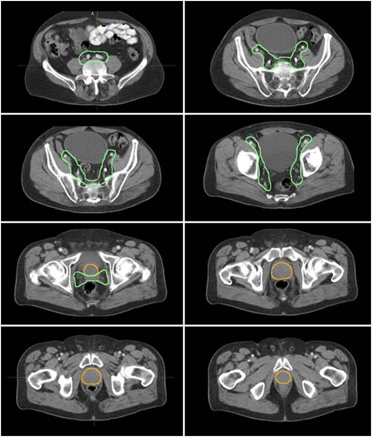
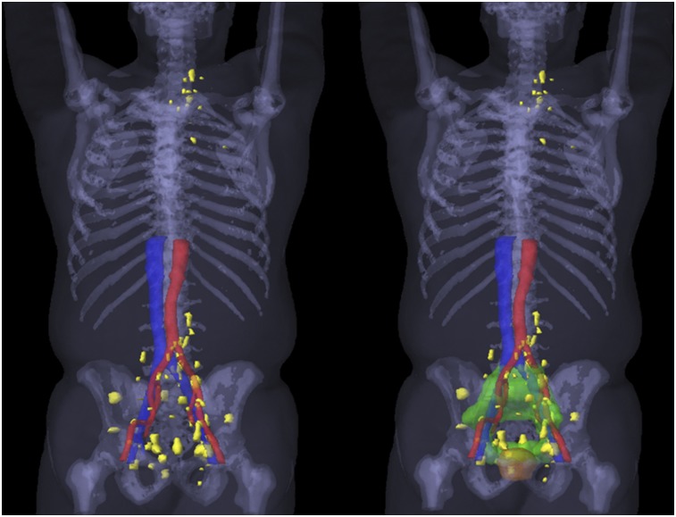
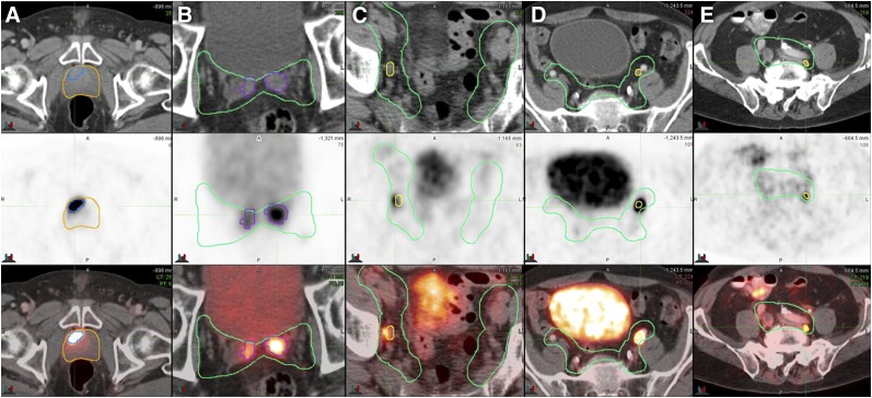
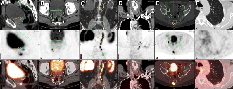
Standard-of-care imaging for initial staging of prostate cancer (PCa) underestimates disease burden. Prostate-specific membrane antigen (PSMA) PET/CT detects PCa metastasis with superior accuracy, having a potential impact on the planning of definitive radiation therapy (RT) for nonmetastatic PCa. Our objectives were to determine how often definitive RT planning based on standard target volumes covers 68Ga-PSMA-11 PET/CT-defined disease and to assess the potential impact of 68Ga-PSMA-11 PET/CT on definitive RT planning. Methods: This was a post hoc analysis of an intention-to-treat population of 73 patients with localized PCa without prior local therapy who underwent 68Ga-PSMA PET/CT for initial staging as part of an investigational new drug trial. Eleven of the 73 were intermediate-risk (15%), 33 were high-risk (45%), 22 were very-high-risk (30%), and 7 were N1 (9.5%). Clinical target volumes (CTVs), which included the prostate, seminal vesicles, and (in accord with the Radiation Therapy Oncology Group consensus guidelines) pelvic lymph nodes (LNs), were contoured on the CT portion of the PET/CT images by a radiation oncologist masked to the PET findings. 68Ga-PSMA-11 PET/CT images were analyzed by a nuclear medicine physician. 68Ga-PSMA-11-positive lesions not covered by planning volumes based on the CTVs were considered to have a major potential impact on treatment planning. Results: All patients had one or more 68Ga-PSMA-11-positive primary prostate lesions. Twenty-five (34%) and 7 (9.5%) of the 73 patients had 68Ga-PSMA-11-positive pelvic LN and distant metastases, respectively. The sites of LN metastases in decreasing order of frequency were external iliac (20.5%), common iliac (13.5%), internal iliac (12.5%) obturator (12.5%), perirectal (4%), abdominal (4%), upper diaphragm (4%), and presacral (1.5%). The median size of the LN lesions was 6 mm (range, 4-24 mm). RT planning based on the CTVs covered 69 (94.5%) of the 73 primary lesions and 20 (80%) of the 25 pelvic LN lesions, on a per-patient analysis. Conclusion:68Ga-PSMA-11 PET/CT had a major impact on intended definitive RT planning for PCa in 12 (16.5%) of the 73 patients whose RT fields covered the prostate, seminal vesicles, and pelvic LNs and in 25 (37%) of the 66 patients whose RT fields covered the prostate and seminal vesicles but not the pelvic LNs.
Keywords: PET/CT; PSMA; definitive radiotherapy; initial staging; prostate cancer.
© 2018 by the Society of Nuclear Medicine and Molecular Imaging.
Figures
References
-
- Alicikus ZA, Yamada Y, Zhang Z, et al. Ten-year outcomes of high-dose, intensity-modulated radiotherapy for localized prostate cancer. Cancer. 2011;117:1429–1437. - PubMed
-
- Sanda MG, Cadeddu JA, Kirkby E, et al. Clinically localized prostate cancer: AUA/ASTRO/SUO guideline. Part I: risk stratification, shared decision making, and care options. J Urol. December 15, 2017. [Epub ahead of print]. - PubMed
-
- Sanda MG, Cadeddu JA, Kirkby E, et al. Clinically localized prostate cancer: AUA/ASTRO/SUO guideline. Part II: recommended approaches and details of specific care options. J Urol. 2018;199:990–997. - PubMed
-
- Lecouvet FE, Geukens D, Stainier A, et al. Magnetic resonance imaging of the axial skeleton for detecting bone metastases in patients with high-risk prostate cancer: diagnostic and cost-effectiveness and comparison with current detection strategies. J Clin Oncol. 2007;25:3281–3287. - PubMed
Publication types
MeSH terms
Substances
Grants and funding
LinkOut - more resources
Full Text Sources
Other Literature Sources
Medical
Miscellaneous