. 2018 May 1;115(18):E4245-E4254.
doi: 10.1073/pnas.1714866115. Epub 2018 Apr 13.

Endoplasmic reticulum stress activates SRC, relocating chaperones to the cell surface where GRP78/CD109 blocks TGF-β signaling

Affiliations

Endoplasmic reticulum stress activates SRC, relocating chaperones to the cell surface where GRP78/CD109 blocks TGF-β signaling

Yuan-Li Tsai et al. Proc Natl Acad Sci U S A. .

Abstract

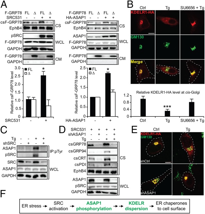
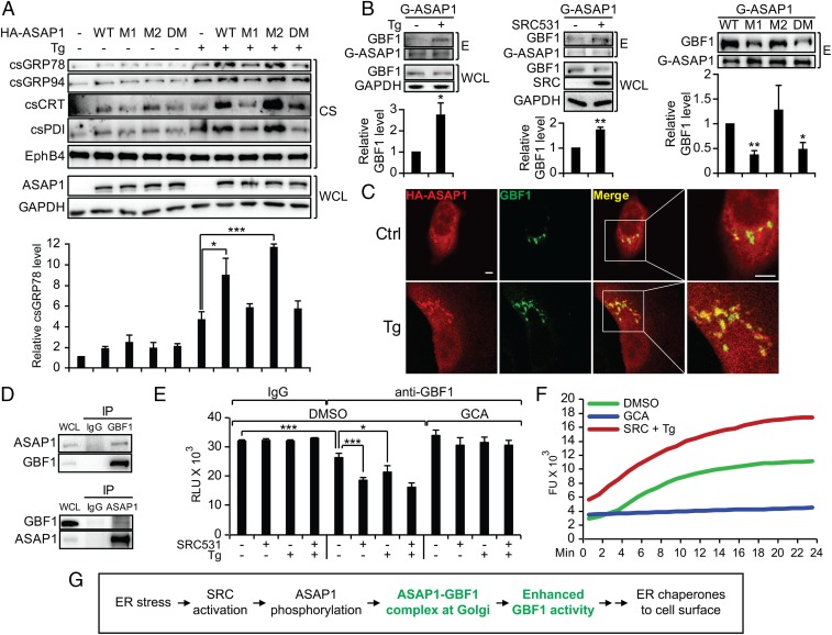
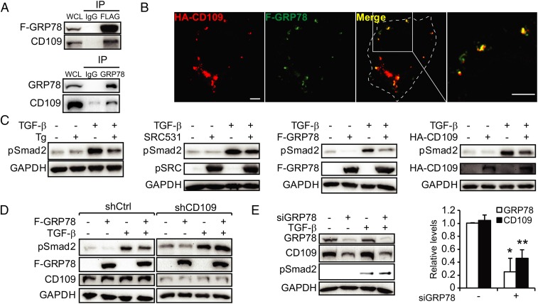
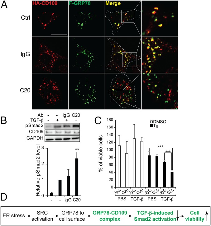
The discovery that endoplasmic reticulum (ER) luminal chaperones such as GRP78/BiP can escape to the cell surface upon ER stress where they regulate cell signaling, proliferation, apoptosis, and immunity represents a paradigm shift. Toward deciphering the mechanisms, we report here that, upon ER stress, IRE1α binds to and triggers tyrosine kinase SRC activation, leading to ASAP1 phosphorylation and Golgi accumulation of ASAP1 and Arf1-GTP, resulting in KDEL receptor dispersion from the Golgi and suppression of retrograde transport. At the cell surface, GRP78 binds to and acts in concert with a glycosylphosphatidylinositol-anchored protein, CD109, in blocking TGF-β signaling by promoting the routing of the TGF-β receptor to the caveolae, thereby disrupting its binding to and activation of Smad2. Collectively, we uncover a SRC-mediated signaling cascade that leads to the relocalization of ER chaperones to the cell surface and a mechanism whereby GRP78 counteracts the tumor-suppressor effect of TGF-β.

Keywords: GRP78; SRC protein kinase; TGF-β signaling; endoplasmic reticulum stress; retrograde transport.


Conflict of interest statement

The authors declare no conflict of interest.

Figures

Fig. 1.
Fig. 1.
IRE1α and SRC regulate cell-surface relocalization of ER chaperones. (A) HeLa cells were treated with Tg for the indicated times. The indicated proteins from whole-cell lysate (WCL) and the cell surface (CS) were analyzed by Western blot with GAPDH and EphB4 serving as loading controls for whole-cell lysate and cell-surface proteins, respectively. In all panels, “pSRC” indicates pSRC(Y419). The band intensities for pSRC and csGRP78 were quantified from three experiments and graphed. (B) As in A, except stable HeLa cell lines expressing shSRC or control shRNA (−) were treated as indicated. (C) As in A, except HeLa cells were transfected with SRC531 or empty vector (−) and were treated as indicated. (D) As in A, except HeLa cells were transfected with IRE1α shRNA (shIRE1α) or control shRNA (−) and were treated with Tg. (E) Western blot analysis of the indicated proteins in HeLa cells with or without Tg treatment. (F) HeLa cells with or without Tg treatment were subjected to co-IP using anti-IRE1α antibody. The indicated proteins were analyzed by Western blot along with whole-cell lysate. (G) IRE1α-knockout HeLa cells expressing HA-IRE1α were treated or were not treated with Tg and were subjected to immunofluorescent staining for the HA epitope (green) and SRC (red). In the merged image, yellow indicates costaining of the two proteins. (Scale bar, 10 µm.) (H) As in F, except IRE1α-knockout HeLa cells transfected with WT HA-IRE1α or the mutant (ΔP) devoid of amino acids 965–977 were not treated or were treated with Tg and subjected to co-IP. (I) Summary of IRE1α and SRC involvement in ER stress-induced chaperone relocalization. *P < 0.05, **P < 0.01, ***P < 0.005.
Fig. 2.
Fig. 2.
The SRC substrate ASAP1 mediates ER stress-induced KDELR1 dispersion and ER chaperone relocalization. (A) HeLa cells were transfected with the full-length F-GRP78 (FL) or the KDEL deletion mutant (Δ) in combination with either SRC531 (Left) or HA-ASAP1 (Right). The indicated proteins from the whole-cell lysate, cell surface, and conditioned medium (CM) were analyzed by Western blot with GAPDH and EphB4 serving as loading controls. The lack of GAPDH in the conditioned medium confirmed cell integrity. The relative csF-GRP78 level under each condition was quantified and graphed. (B) HeLa cells were transfected with KDELR1-HA, pretreated with SU6656 followed by Tg, and subjected to immunofluorescent (IF) staining for the HA epitope (red) and GM130 (green), with the latter serving as marker for cis-Golgi. In the merged image, yellow indicates costaining of the two proteins. The white dashed lines outline the cell shape. (Scale bar, 10 µm.) The immunofluorescent intensities of KDELR1-HA at the cis-Golgi were quantified and graphed. (C) HeLa cells expressing shSRC or control shRNA (−) with or without Tg treatment were subjected to immunoprecipitation (IP) using anti–phospho-tyrosine (pTyr) antibody. The indicated proteins were analyzed by Western blot along with whole-cell lysate. (D) As in A, except HeLa cells expressing shASAP1 or control shRNA (−) transfected with the SRC531 expression vector were treated as indicated. (E) As in B, except HeLa cells expressing shASAP1 or control shRNA (shCtrl) were treated as indicated. (Scale bar, 5 µm.) (F) Summary of ASAP1 and KDELR1 involvement in ER stress-induced chaperone relocalization. *P < 0.05; ***P < 0.005.
Fig. 3.
Fig. 3.
ER stress promotes ASAP1 and GBF1 complex formation and enhances GBF1 GEF activity. (A) HeLa cells transfected with HA-ASAP1 WT or mutant expression vectors (M1, M2, and DM) were not treated or were treated with Tg. The indicated proteins from whole-cell lysate or the cell-surface preparations were analyzed by Western blots with GAPDH and EphB4 serving as loading controls. The csGRP78 levels were quantified and graphed. (B) HeLa cells were treated with Tg (Left) or were transfected with SRC531 expression vector (Middle) or both (Right). Whole-cell lysate was subjected to a pull-down assay with GST-fused WT or mutant ASAP1 recombinant proteins (G-ASAP1). The indicated proteins from the eluate (E) were analyzed by Western blot. The relative eluted GBF1 level under each condition was quantified and graphed with SD. (C) HeLa cells transfected with the HA-ASAP1 expression vector were not treated (Ctrl) or were treated with Tg and were subjected to immunofluorescent staining for the HA epitope (red) or GBF-1 (green). Costaining of both proteins (yellow) is shown in the merged images and the enlarged views. (Scale bars, 5 µm.) (D) HeLa cells were transfected with the SRC531 expression vector followed by Tg treatment, and the whole-cell lysate was subjected to immunoprecipitation using IgG control, anti-GBF1, or anti-ASAP1 antibodies. The indicated proteins were analyzed by Western blot. (E) HeLa cells were transfected with the SRC531 expression vector and were treated with Tg, DMSO, or GCA as indicated. The whole-cell lysates were subjected to immunoprecipitation using anti-GBF1 or control IgG antibodies, and the immunoprecipitate was subjected to the GTPase-Glo GEF activity assay. (F) As in E, except the GBF1 immunoprecipitates from the indicated treatment conditions were subjected to the GTP exchange assay. (G) Summary of ASAP1 and GBF1 involvement in the ER stress-induced chaperone relocalization. *P < 0.05, **P < 0.01, ***P < 0.005.
Fig. 4.
Fig. 4.
GBF1 facilitates the ER stress-induced Arf1-GTP increase at the cis-Golgi. (A) The indicated cancer cell lines were treated with Tg and GCA as indicated. Cell-surface proteins were isolated and probed for the indicated proteins by Western blot with EphB4 serving as loading control. (B) Whole-cell lysate from HeLa cells treated with Tg or transfected with SRC531 or HA-ASAP1 expression vectors were subjected to the Arf1 pull-down activation assay. The indicated proteins from the eluate (E) or whole-cell lysate were analyzed by Western blot. The band intensities for Arf1-GTP were quantified and graphed. (C) As in B, except the cells were treated additionally with GCA as indicated. (D) As in B, except SRC (shSRC)- or ASAP1 (shASAP1)-knockdown stable HeLa cell lines were treated with Tg as indicated. (E) SK-MEL-28 cells were transfected with either WT Arf1-HA or the Q71L mutant expression vectors. The cells were not treated (Ctrl), treated with Tg, or pretreated with SU6656 followed by Tg and were subjected to immunofluorescent staining for the HA epitope (red) or GM130 (green). Yellow indicates costaining of the two proteins in the merged images. The immunofluorescent intensities of Arf1-HA at the cis-Golgi under each condition were quantified and graphed. (Scale bar, 10 µm.) *P < 0.05, ***P < 0.005.
Fig. 5.
Fig. 5.
GTP-locked Arf1(Q71L) is sufficient to drive KDELR1 dispersion and ER chaperones to the cell surface. (A) The indicated cancer cell lines were transfected with the Arf1-HA(Q71L) mutant expression vector and were treated with Tg as indicated. The indicated proteins from the whole-cell lysate and the cell surface were analyzed by Western blot with GAPDH and EphB4 serving as loading controls. (B) As in A, except HeLa cells were transfected with the indicated expression vectors, and conditioned medium was collected and assayed for secreted F-GRP78. The band intensities of csF-GRP78 were quantified and graphed. (C) HeLa cells were transfected with the KDELR1-FLAG (KDELR1-F) expression vector alone or in combination with expression vectors for SRC531-His, HA-ASAP1, or Arf1-HA(Q71L), as indicated, and were subjected to immunofluorescent staining for the indicated proteins. GM130 served as the marker for cis-Golgi. (Scale bar, 5 µm.) (D) Summary of the ER stress-signaling cascade leading to the escape of ER luminal chaperones to the cell surface. *P < 0.05.
Fig. 6.
Fig. 6.
csGRP78 partners with CD109 in TGF-β inhibition. (A) Whole-cell lysate from HeLa cells expressing F-GRP78 (Upper) or SK-MEL-28 cells (Lower) were subjected to immunoprecipitation with IgG, anti-FLAG, or anti-GRP78 antibodies as indicated. The immunoprecipitate along with the whole-cell lysate was probed by Western blot for F-GRP78, GRP78, and CD109. (B) HeLa cells were cotransfected with F-GRP78 and HA-CD109 expression vectors and were subjected to immunofluorescent staining for the HA (red) and FLAG (green) epitopes. Costaining of the two proteins is indicated by yellow in the merged image and the enlarged view. (Scale bars, 5 µm.) (C) HeLa cells were treated with Tg or TGF-β and were transfected with SRC531, F-GRP78, or HA-CD109 alone or in combination, as indicated. The indicated proteins were analyzed by Western blot. (D) As in C, except the CD109 (shCD109)- or control shRNA (shCtrl)-knockdown stable HeLa cell line was treated as indicated. (E) As in D, except SK-MEL-28 cells were transfected with siRNA against GRP78 (siGRP78) and were treated with TGF-β as indicated. The band intensities were quantified and graphed. *P < 0.05, **P < 0.01.
Fig. 7.
Fig. 7.
csGRP78 promotes the routing of TβR1 to the caveolae and disrupts its interaction with Smad2. (A) SK-MEL-28 cells transfected with TβR1-His or F-GRP78, alone or in combination were treated with TGF-β as indicated. The cells were subjected to coimmunoprecipitation using anti-His antibody. The indicated proteins were analyzed by Western blot. (B) SK-MEL-28 cells were transfected with pcDNA3 or F-GRP78 or were treated with TGF-β alone or in combination as indicated. The whole-cell lysate was subjected to sucrose-gradient fractionation. The fractions were subjected to Western blot for analysis of the indicated proteins. CAV-1, caveolin 1. The lipid raft was enriched in fraction 5, and nonraft was enriched in fractions 9–12 with CAV-1 and GAPDH serving as markers for the respective fractions. The percentage of TβR1 in each fraction was quantified and graphed. (C) Model of the inhibition of TGF-β signaling by csGRP78/CD109. Upon TGF-β stimulation, csGRP78/CD109 routes the TβR to the caveolae for degradation, disrupting the binding of the receptor to Smad2 and its subsequent activation, thereby blunting TGF-β–mediated growth inhibition.
Fig. 8.
Fig. 8.
Effect of antibody targeting GRP78 on the interaction with CD109 and TGF-β signaling. (A) HeLa cells cotransfected with F-GRP78 and HA-CD109 were not treated (Ctrl) or were treated with IgG or the anti-GRP78 antibody C20. The nonpermeabilized cells were subjected to immunofluorescent staining for the HA (red) and FLAG (green) epitopes. The yellow staining in the merged images and enlarged views indicate costaining of the csF-GRP78 with HA-CD109, which was disrupted by C20 treatment. (Scale bar, 10 µm.) (B) SK-MEL-28 cells were treated with IgG, C20, and TGF-β as indicated. The indicated proteins were analyzed by Western blot. The band intensities of pSmad2 were quantified and graphed. (C) As in B, except the cells were treated with DMSO or Tg in combination with the antibodies and then were treated with PBS or TGF-β and subjected to the WST-1 assay. (D) Summary of GRP78/CD109 inhibition of TGF-β signaling and promotion of survival under ER stress. **P < 0.01, ***P < 0.005.

Similar articles

Cited by

References

    1. Shiu RP, Pouysségur J, Pastan I. Glucose depletion accounts for the induction of two transformation-sensitive membrane proteinsin Rous sarcoma virus-transformed chick embryo fibroblasts. Proc Natl Acad Sci USA. 1977;74:3840–3844. - PMC - PubMed
    1. Ni M, Zhang Y, Lee AS. Beyond the endoplasmic reticulum: Atypical GRP78 in cell viability, signalling and therapeutic targeting. Biochem J. 2011;434:181–188. - PMC - PubMed
    1. Behnke J, Mann MJ, Scruggs FL, Feige MJ, Hendershot LM. Members of the Hsp70 family recognize distinct types of sequences to execute ER quality control. Mol Cell. 2016;63:739–752. - PMC - PubMed
    1. Lee AS. Glucose-regulated proteins in cancer: Molecular mechanisms and therapeutic potential. Nat Rev Cancer. 2014;14:263–276. - PMC - PubMed
    1. Zhang Y, Liu R, Ni M, Gill P, Lee AS. Cell surface relocalization of the endoplasmic reticulum chaperone and unfolded protein response regulator GRP78/BiP. J Biol Chem. 2010;285:15065–15075. - PMC - PubMed

Publication types

MeSH terms