. 2018 May 1;115(18):4749-4754.
doi: 10.1073/pnas.1718217115. Epub 2018 Apr 13.

Role of PD-1 during effector CD8 T cell differentiation

Affiliations

Role of PD-1 during effector CD8 T cell differentiation

Eunseon Ahn et al. Proc Natl Acad Sci U S A. .

Abstract

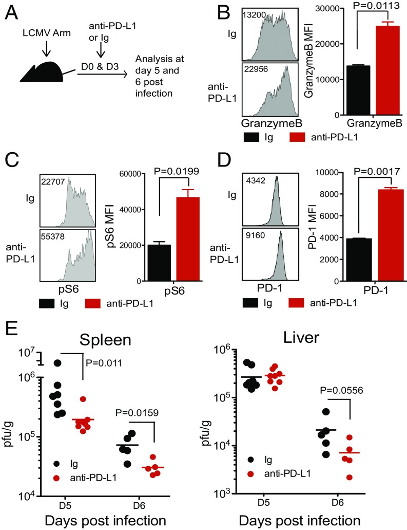
PD-1 (programmed cell death-1) is the central inhibitory receptor regulating CD8 T cell exhaustion during chronic viral infection and cancer. Interestingly, PD-1 is also expressed transiently by activated CD8 T cells during acute viral infection, but the role of PD-1 in modulating T cell effector differentiation and function is not well defined. To address this question, we examined the expression kinetics and role of PD-1 during acute lymphocytic choriomeningitis virus (LCMV) infection of mice. PD-1 was rapidly up-regulated in vivo upon activation of naive virus-specific CD8 T cells within 24 h after LCMV infection and in less than 4 h after peptide injection, well before any cell division had occurred. This rapid PD-1 expression by CD8 T cells was driven predominantly by antigen receptor signaling since infection with a LCMV strain with a mutation in the CD8 T cell epitope did not result in the increase of PD-1 on antigen-specific CD8 T cells. Blockade of the PD-1 pathway using anti-PD-L1 or anti-PD-1 antibodies during the early phase of acute LCMV infection increased mTOR signaling and granzyme B expression in virus-specific CD8 T cells and resulted in faster clearance of the infection. These results show that PD-1 plays an inhibitory role during the naive-to-effector CD8 T cell transition and that the PD-1 pathway can also be modulated at this stage of T cell differentiation. These findings have implications for developing therapeutic vaccination strategies in combination with PD-1 blockade.

Keywords: CD8 T cells; PD-1; effector differentiation; memory cells; viral infection.


Conflict of interest statement

Conflict of interest statement: R.A. has patents on the PD-1 pathway and has served on the advisory board for Genentech/Roche and received research funding from Genentech and Merck. G.J.F. and A.H.S. have patents/pending royalties on the PD-1 pathway from Roche, Merck, Bristol-Myers Squibb, EMD Serono, Boehringer Ingelheim, AstraZeneca, Dako, and Novartis. G.J.F. has served on advisory boards for Roche, Bristol-Myers Squibb, Xios, and Quiet. A.H.S. has served on advisory boards for Novartis, Surface Oncology, and Elstar. A.H.S. has received research funding from Novartis, Roche, UCB, Ipsen, and Quark. B.I.A. is an employee of Five Prime Therapeutics.

Figures

Fig. 1.
Fig. 1.
PD-1 is rapidly expressed by antigen-specific CD8 T cells after viral infection. (A) The 1 × 10⁶ CellTrace Violet (CTV)-labeled naive P14 TCR transgenic CD8 T cells (LCMV gp33-specific) were adoptively transferred into B6 mice. These P14 chimeric mice were infected with LCMV Arm (2 × 10⁶ pfu/mouse i.v.). (B) Flow plots were gated on P14 cells. Expression of PD-1 and other activation markers (CD44, CD25, and CD69) was analyzed in the spleen at days 1, 2, and 3 post infection. (C) PD-1 expression on P14 cells was compared in mice infected with different doses of LCMV Arm (2 × 10², 2 × 10⁴, or 2 × 10⁶ pfu/mouse i.v.) at days 1, 2, and 3 post infection. Data are representative of three independent experiments with three to four mice per group.
Fig. 2.
Fig. 2.
Expression kinetics of inhibitory receptors and effector molecules on virus-specific CD8 T cells during early phase of T cell activation. CellTrace Violet-labeled P14 cells were transferred into B6 mice, followed by LCMV Arm infection (2 × 10⁶ pfu/mouse i.v.). (A) CTLA-4, Tim-3, LAG-3, and 2B4 expression on P14 cells were examined at days 1, 2, and 3 post infection. (B) Granzyme B expression on P14 cells was observed at days 1, 2, and 3 post infection. To analyze IFNγ and TNFα production, splenocytes from P14 chimeric mice were stimulated with GP33 peptide for 5 h at days 1, 2, and 3 post infection. Data are representative of two independent experiments with three mice per group.
Fig. 3.
Fig. 3.
TCR signal is the primary regulator of PD-1 expression. (A) P14 chimeric mice were infected with either WT LCMV or a LCMV strain with a mutation (V35A) in the GP33 epitope. PD-1 expression was examined on P14 cells in the spleen at days 1, 2, and 3 post infection. (B) After LCMV infection (2 × 10⁶ pfu/mouse i.v.), P14 chimeric mice were treated with FK506 at day 3 post infection. PD-1 expression was examined in P14 cells at days 4 and 5 post infection. Data are representative of two to three independent experiments with three to five mice per group.
Fig. 4.
Fig. 4.
Naive antigen-specific CD8 T cells express PD-1 within 4 h after peptide immunization. (A) GP33-41 peptide (200 μg/mouse i.v.) was injected into P14 chimeric mice, and PD-1, CD25, and CD69 expression on P14 cells was examined at 2, 4, 8, and 24 h post peptide injection. (B) Six hours after peptide injection, P14 chimeric mice were treated with FK506, and PD-1 expression was examined in P14 cells at 24 h post peptide injection. Data are representative of three independent experiments with three mice per group.
Fig. 5.
Fig. 5.
PD-1 pathway blockade during early phase of viral infection enhances effector function of antigen-specific CD8 T cells and reduces viral load. LCMV-infected mice were treated with anti–PD-L1 antibody (10F.9G2) or control Ig at days 0 and 3 post infection. (A) Experimental setup. (B) Granzyme B expression by DbGP33+ CD8 T cells at day 5 post infection. (C) Phosphorylation of S6 on P14 cells was examined at day 5 post infection. Representative flow plots and summary graphs on DbGP33+ CD8 T cells and P14 cells are shown. (D) PD-1 expression on DbGP33+ CD8 T cells at day 5. (E) Viral titers in spleen and liver were examined at days 5 and 6 post infection. Data are representative of two to three independent experiments with three to five mice per group. Error bars represent the SEM.
Fig. 6.
Fig. 6.
PD-1 blockade during acute viral infection enhances the frequency of the memory precursor subset of effector CD8 T cells. (A) Mice were treated with anti–PD-1(29F10) antibody or control Ig at days 0, 3, 6, and 9 post LCMV Arm. (B) The number of GP33+ CD8 T cells in PBMCs was analyzed at days 8, 15, and 30 post infection. (C) Gated on GP33+ CD8 T cells, the MP subset (CD127hi KLRG1low) and the TE subset (CD127low KLRG1hi) were analyzed at days 8, 15, and 30 post infection. (Right) Summary graph. (Bottom) CD62L expression in DbGP33+ CD8 T cells was examined at days 8, 15, and 30 post infection. (Right) Summary graph. (D) Mice were treated with anti–PD-1(29F10) antibody or control Ig at days 0, 3, 6, and 9 post Ad5-GP immunization. (E) The number of GP33+ CD8 T cells in PBMCs was analyzed at day 8, 15, and 22 post immunization. (F) Gated on GP33+ CD8 T cells, MP and TE subsets were analyzed at days 8, 15, and 22 post immunization. Summary graph is shown at Right. Data are representative of three independent experiments with five mice per group. Error bars represent the SEM.

References

    1. Ahmed R, Gray D. Immunological memory and protective immunity: Understanding their relation. Science. 1996;272:54–60. - PubMed
    1. Obar JJ, Lefrançois L. Memory CD8+ T cell differentiation. Ann N Y Acad Sci. 2010;1183:251–266. - PMC - PubMed
    1. Akondy RS, et al. Origin and differentiation of human memory CD8 T cells after vaccination. Nature. 2017;552:362–367. - PMC - PubMed
    1. Kaech SM, et al. Selective expression of the interleukin 7 receptor identifies effector CD8 T cells that give rise to long-lived memory cells. Nat Immunol. 2003;4:1191–1198. - PubMed
    1. Kaech SM, Wherry EJ, Ahmed R. Effector and memory T-cell differentiation: Implications for vaccine development. Nat Rev Immunol. 2002;2:251–262. - PubMed

Publication types

MeSH terms

Substances