Review
. 2018 Apr 13;131(8):jcs189928.
doi: 10.1242/jcs.189928.

Amyloid assembly and disassembly

Affiliations
Review

Amyloid assembly and disassembly

Edward Chuang et al. J Cell Sci. .

Abstract

Amyloid fibrils are protein homopolymers that adopt diverse cross-β conformations. Some amyloid fibrils are associated with the pathogenesis of devastating neurodegenerative disorders, including Alzheimer's disease and Parkinson's disease. Conversely, functional amyloids play beneficial roles in melanosome biogenesis, long-term memory formation and release of peptide hormones. Here, we showcase advances in our understanding of amyloid assembly and structure, and how distinct amyloid strains formed by the same protein can cause distinct neurodegenerative diseases. We discuss how mutant steric zippers promote deleterious amyloidogenesis and aberrant liquid-to-gel phase transitions. We also highlight effective strategies to combat amyloidogenesis and related toxicity, including: (1) small-molecule drugs (e.g. tafamidis) to inhibit amyloid formation or (2) stimulate amyloid degradation by the proteasome and autophagy, and (3) protein disaggregases that disassemble toxic amyloid and soluble oligomers. We anticipate that these advances will inspire therapeutics for several fatal neurodegenerative diseases.

Keywords: Amyloid; Autophagy; Disaggregase; Neurodegeneration; Prion.


Conflict of interest statement

Competing interestsThe authors declare no competing or financial interests.

Figures
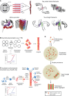
Fig. 1.
Fig. 1.
Amyloid structure and formation pathways. (A) Top-left: the X-ray diffraction pattern for amyloids shows major reflections at ∼4.7 Å (hydrogen bonding distances between β-strands) and ∼10 Å (side-chain packing between β-sheets) indicating cross-β structure where β-strands align perpendicular to the fibril axis. Bottom-left: solid-state NMR structure of human α-synuclein fibril (PDB: 2N0A) (Tuttle et al., 2016). Right side: 3.4 Å–3.5 Å resolution cryo-EM structures of tau paired-helical filaments (PDB: 5O3L) and straight filaments (PDB: 5O3T) from an AD patient (Fitzpatrick et al., 2017). (B) In downhill polymerization (DP), the lag phase of amyloid formation is due to the slow dissociation of a stable native tetramer into monomers, which then rapidly assume an amyloidogenic conformation. This mechanism is employed by TTR in FAP (Hurshman et al., 2004). TTR amyloidosis can be inhibited by tafamidis, a drug that stabilizes TTR in its native tetrameric state (Coelho et al., 2012). Thus, understanding the mechanism of amyloid formation can enable development of drugs to preserve the native state and prevent amyloidogenesis. Typically, amyloids formed by DP do not eliminate the lag phase of fibrillization in reactions seeded with preformed fibrils (lower panel). (C) In nucleated conformational conversion (NCC), partially or completely disordered soluble monomers are initially in equilibrium with molten soluble oligomers. During the lag phase of assembly, these molten soluble oligomers gradually rearrange into amyloidogenic oligomers, which then rapidly form cross-β nuclei (primary nucleation), thereby ending the lag phase. As soon as cross-β nuclei have formed, fibrillization proceeds rapidly as nuclei recruit and convert soluble monomers and molten soluble oligomers into the cross-β form at the growing fibril ends. The introduction of pre-formed fibrils eliminates the lag phase of assembly via immediate templating of the amyloid conformation. The lateral face of the assembled fibril also serves as a site for secondary nucleation events where molten oligomers or soluble monomers can rapidly convert into amyloidogenic oligomers. Typically, amyloids formed by NCC eliminate the lag phase of fibrillization in reactions seeded with preformed fibrils (lower panel). (D) Phase transition of proteins containing prion-like domains (PrLDs). RBPs can condense into liquid droplets through transient interactions between PrLDs and other multivalent interactions. Droplet persistence enables formation of stable (less dynamic) interactions between PrLDs that drive an aberrant phase transition from liquid to solid states that comprise pathological fibrils, which accumulate in disease.
Fig. 2.
Fig. 2.
Functional amyloids. (A) PMEL forms functional amyloid in melanin metabolism. PMEL fibril formation is highly regulated by post-translational cleavage into its amyloidgenic form and compartmentalization within melanosomes during melanosome maturation. PMEL fibrils catalyze the formation of melanin, concentrate melanin and facilitate bulk transport of melanin (Watt et al., 2013). (B) CPEB3 is a regulator of mRNA translation in neurons and enhances LTP through positive regulation of AMPA receptor translation. CPEB3 is soluble and SUMOylated in its basal state. Upon neuronal activation, CPEB3 is deSUMOylated and ubiquitylated, causing the protein to aggregate and activate translation of certain mRNAs (Drisaldi et al., 2015). (C) Peptide hormones (blue) are concentrated in secretory granules where they form amyloids (red) as a packaging mechanism. Some peptide hormones aggregate spontaneously, while others require the assistance of glycosaminoglycans (Maji et al., 2009). Furthermore, these amyloid fibrils slowly depolymerize spontaneously upon vesicle release into the extracellular space, resulting in delayed release of monomeric hormones.
Fig. 3.
Fig. 3.
Amyloid degradation via autophagy and the ubiquitin-proteasome system. (A) In macroautophagy, K63 poly-ubiquitylated aggregates are engulfed by autophagosomes and targeted for degradation. Fusion of the autophagosome with a lysosome forms an autolysosome that degrades the aggregate cargo. Lysosome acidification relies on presenilin 1 (PS1), which recruits a proton pump to the lysosome that is critical for autolysosome acidification (denoted *) (Lee et al., 2010b). (B) In neurons, autophagosome formation occurs in the distal axon. Autophagosomes then fuse with late endosomes as they are retrogradely transported along microtubules by dynein toward the soma. Autophagosomes also bind kinesin motors, which must be negatively regulated to yield robust retrograde motility driven by dynein. Upon arrival in the soma, autophagosomes mature into autolysosomes via fusion with lysosomes. (C) Protein disaggregases such as Hsp70 in combination with Hsp110 and Hsp40 can extract polypeptides from aggregates and allow them to: (1) refold, (2) be degraded by the proteasome or (3) be degraded by chaperone-mediated autophagy. Polypeptides extracted from aggregates can be ubiquitylated by Hsp70-associated ubiquitin ligases such as CHIP (McDonough and Patterson, 2003). The polypeptides are then brought to the proteasome for degradation by shuttles such as UBQLN2 (Hjerpe et al., 2016). Tau fibrils can inhibit proteasome activity and this inhibition can be relieved by increasing cAMP–PKA signaling with the small-molecule Rolipram (Myeku et al., 2016). Alternatively, polypeptides may be preferentially translocated into the lysosome for degradation via a process called chaperone-mediated autophagy (Schneider and Cuervo, 2013).
Fig. 4.
Fig. 4.
Amyloid-disaggregase machineries. (A) Hsp104 is an AAA+ ATPase with the ability to efficiently fragment yeast prions to allow their inheritance by daughter cells. Hsp104 can fragment amyloid fibrils by partial or full translocation of a polypeptide out of the fibril, thus creating a break point (Sweeny and Shorter, 2016). (B) Hsp70 family proteins contain a nucleotide-binding domain and a substrate-binding domain. Polypeptides trapped in fibrils are recruited to the substrate-binding domain of Hsp70 by Hsp40 family proteins. Concomitant binding of Hsp40 and substrate to Hsp70 facilitates ATP hydrolysis and a conformational change in Hsp70 to a closed state, which traps the substrate. Then through a poorly understood mechanism, in conjunction with Hsp110 family proteins, nucleotide exchange factors for Hsp70, polypeptide is extracted and refolded into its native conformation (Nillegoda and Bukau, 2015; Torrente and Shorter, 2013). This process may require Hsp110 to engage substrate and hydrolyze ATP (Mattoo et al., 2013; Scior et al., 2018; Shorter, 2011). Hsp110, Hsp70 and Hsp40 preferentially depolymerize amyloid fibrils from their ends (Duennwald et al., 2012; Gao et al., 2015). (C) Human HtrA1 is an ATP-independent serine protease that functions as a homotrimer. HtrA1 has the PDZ domain-dependent ability to disassemble Aβ and tau fibrils followed by subsequent proteolysis by its serine protease domain (Poepsel et al., 2015).

References

    1. Abedini A., Plesner A., Cao P., Ridgway Z., Zhang J., Tu L.-H., Middleton C. T., Chao B., Sartori D. J., Meng F. et al. (2016). Time-resolved studies define the nature of toxic IAPP intermediates, providing insight for anti-amyloidosis therapeutics. 5, e12977 10.7554/eLife.12977 - DOI - PMC - PubMed
    1. Abisambra J., Jinwal U. K., Miyata Y., Rogers J., Blair L., Li X., Seguin S. P., Wang L., Jin Y., Bacon J. et al. (2013). Allosteric heat shock protein 70 inhibitors rapidly rescue synaptic plasticity deficits by reducing aberrant tau. 74, 367-374. 10.1016/j.biopsych.2013.02.027 - DOI - PMC - PubMed
    1. Aguzzi A. and Calella A. M. (2009). Prions: protein aggregation and infectious diseases. 89, 1105-1152. 10.1152/physrev.00006.2009 - DOI - PubMed
    1. Alami N. H., Smith R. B., Carrasco M. A., Williams L. A., Winborn C. S., Han S. S. W., Kiskinis E., Winborn B., Freibaum B. D., Kanagaraj A. et al. (2014). Axonal transport of TDP-43 mRNA granules is impaired by ALS-causing mutations. 81, 536-543. 10.1016/j.neuron.2013.12.018 - DOI - PMC - PubMed
    1. Alberti S., Halfmann R., King O., Kapila A. and Lindquist S. (2009). A systematic survey identifies prions and illuminates sequence features of prionogenic proteins. 137, 146-158. 10.1016/j.cell.2009.02.044 - DOI - PMC - PubMed

Publication types

LinkOut - more resources