

Proteomic analysis at the sites of clinical infection with invasive Streptococcus pyogenes
- PMID: 29654237
- PMCID: PMC5899161
- DOI: 10.1038/s41598-018-24216-2
Proteomic analysis at the sites of clinical infection with invasive Streptococcus pyogenes
Abstract
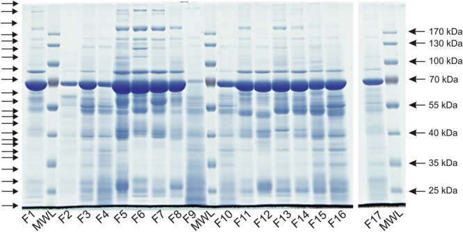
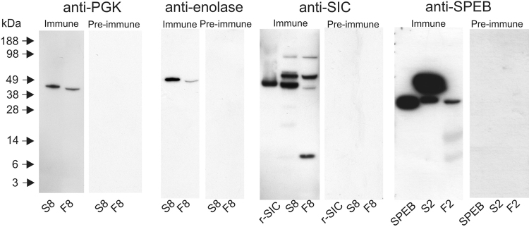
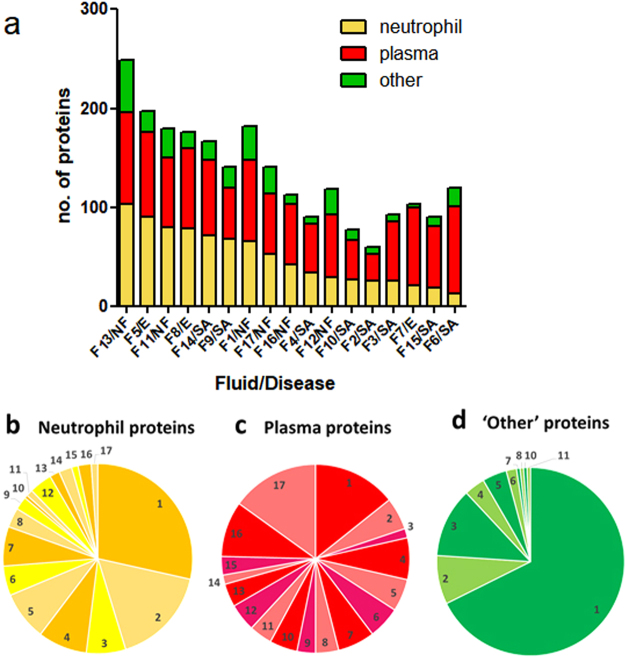
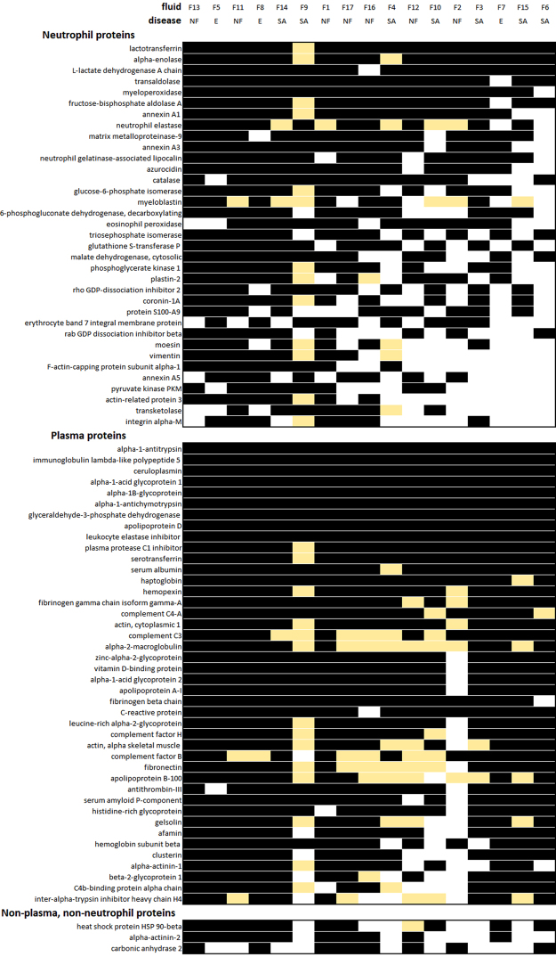
Invasive Streptococcus pyogenes infections are rare, with often-unexplained severity. Prompt diagnosis is desirable, as deaths can occur rapidly following onset and there is an increased, but preventable, risk to contacts. Here, proteomic analyses of clinical samples from invasive human S. pyogenes infections were undertaken to determine if novel diagnostic targets could be detected, and to augment our understanding of disease pathogenesis. Fluid samples from 17 patients with confirmed invasive S. pyogenes infection (empyema, septic arthritis, necrotising fasciitis) were analysed by proteomics for streptococcal and human proteins; 16/17 samples had detectable S. pyogenes DNA. Nineteen unique S. pyogenes proteins were identified in just 6/17 samples, and 15 of these were found in a single pleural fluid sample including streptococcal inhibitor of complement, trigger factor, and phosphoglycerate kinase. In contrast, 469 human proteins were detected in patient fluids, 177 (38%) of which could be identified as neutrophil proteins, including alpha enolase and lactotransferrin which, together, were found in all 17 samples. Our data suggest that streptococcal proteins are difficult to detect in infected fluid samples. A vast array of human proteins associated with leukocyte activity are, however, present in samples that deserve further evaluation as potential biomarkers of infection.
Conflict of interest statement
The authors declare no competing interests.
Figures
Similar articles
-
Label-free proteomic analysis of environmental acidification-influenced Streptococcus pyogenes secretome reveals a novel acid-induced protein histidine triad protein A (HtpA) involved in necrotizing fasciitis.J Proteomics. 2014 Sep 23;109:90-103. doi: 10.1016/j.jprot.2014.06.026. Epub 2014 Jul 3. J Proteomics. 2014. PMID: 24998435
-
Streptococcus pyogenes Transcriptome Changes in the Inflammatory Environment of Necrotizing Fasciitis.Appl Environ Microbiol. 2019 Oct 16;85(21):e01428-19. doi: 10.1128/AEM.01428-19. Print 2019 Nov 1. Appl Environ Microbiol. 2019. PMID: 31471300 Free PMC article.
-
A comprehensive analysis of the Streptococcus pyogenes and human plasma protein interaction network.Mol Biosyst. 2014 Jul;10(7):1698-708. doi: 10.1039/c3mb70555b. Epub 2014 Feb 14. Mol Biosyst. 2014. PMID: 24525632
-
Intracellular Invasion by Streptococcus pyogenes: Invasins, Host Receptors, and Relevance to Human Disease.Microbiol Spectr. 2019 Jul;7(4):10.1128/microbiolspec.gpp3-0049-2018. doi: 10.1128/microbiolspec.GPP3-0049-2018. Microbiol Spectr. 2019. PMID: 31267891 Free PMC article. Review.
-
Severe invasive streptococcal infection by Streptococcus pyogenes and Streptococcus dysgalactiae subsp. equisimilis.Microbiol Immunol. 2016 Jan;60(1):1-9. doi: 10.1111/1348-0421.12334. Microbiol Immunol. 2016. PMID: 26762200 Review.
Cited by
-
Vaccine-induced, but not natural immunity, against the Streptococcal inhibitor of complement protects against invasive disease.NPJ Vaccines. 2021 Apr 22;6(1):62. doi: 10.1038/s41541-021-00326-3. NPJ Vaccines. 2021. PMID: 33888727 Free PMC article.
-
A model for predicting bacteremia species based on host immune response.Front Cell Infect Microbiol. 2025 Feb 18;15:1451293. doi: 10.3389/fcimb.2025.1451293. eCollection 2025. Front Cell Infect Microbiol. 2025. PMID: 40041147 Free PMC article.
-
Streptococcus pyogenes emm98.1 variants activate inflammatory caspases in human neutrophils.Virulence. 2023 Dec;14(1):2264090. doi: 10.1080/21505594.2023.2264090. Epub 2023 Oct 13. Virulence. 2023. PMID: 37830540 Free PMC article.
-
Dual-species proteomics and targeted intervention of animal-pathogen interactions.J Adv Res. 2025 Jul;73:397-410. doi: 10.1016/j.jare.2024.08.038. Epub 2024 Sep 2. J Adv Res. 2025. PMID: 39233003 Free PMC article.
-
Extracellular bacterial lymphatic metastasis drives Streptococcus pyogenes systemic infection.Nat Commun. 2020 Sep 17;11(1):4697. doi: 10.1038/s41467-020-18454-0. Nat Commun. 2020. PMID: 32943639 Free PMC article.
References
-
- Stirling, P., Tahir, M. & Atkinson, H. D. The limitations of Gram-stain microscopy of synovial fluid in concomitant septic and crystal arthritis. Curr. Rheumatol. Rev. 13, 10.2174/1573397113666170329123308 (2017). - PubMed
Publication types
MeSH terms
Substances
Grants and funding
LinkOut - more resources
Full Text Sources
Other Literature Sources
Medical
