. 2018 Apr 13;8(1):5975.
doi: 10.1038/s41598-018-24199-0.

Defective immuno- and thymoproteasome assembly causes severe immunodeficiency

Affiliations

Defective immuno- and thymoproteasome assembly causes severe immunodeficiency

Irina Treise et al. Sci Rep. .

Abstract

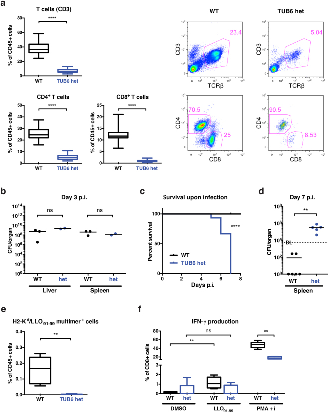
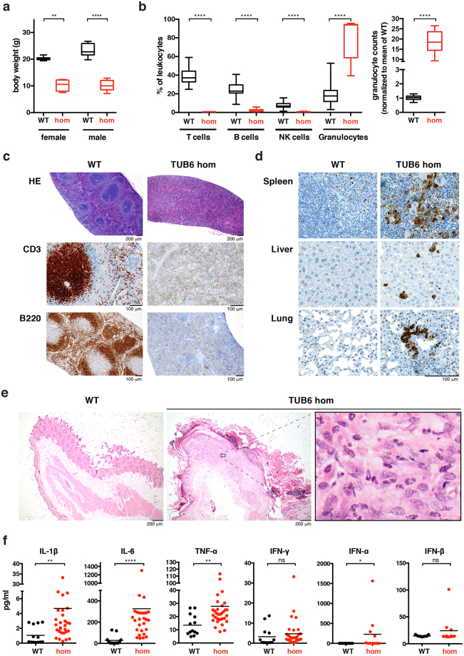
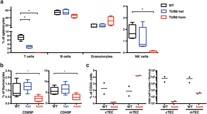
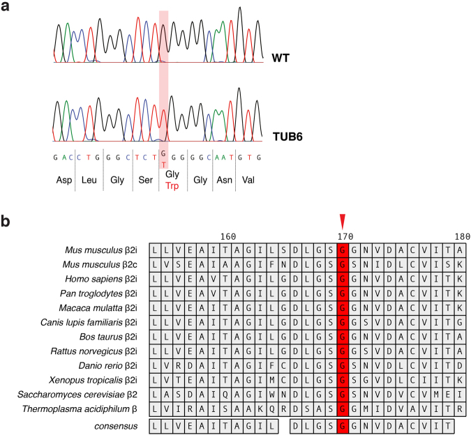
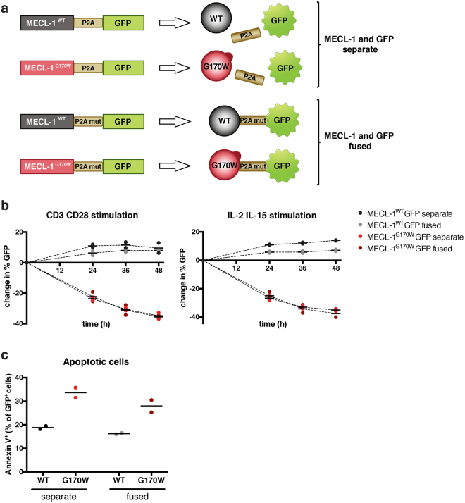
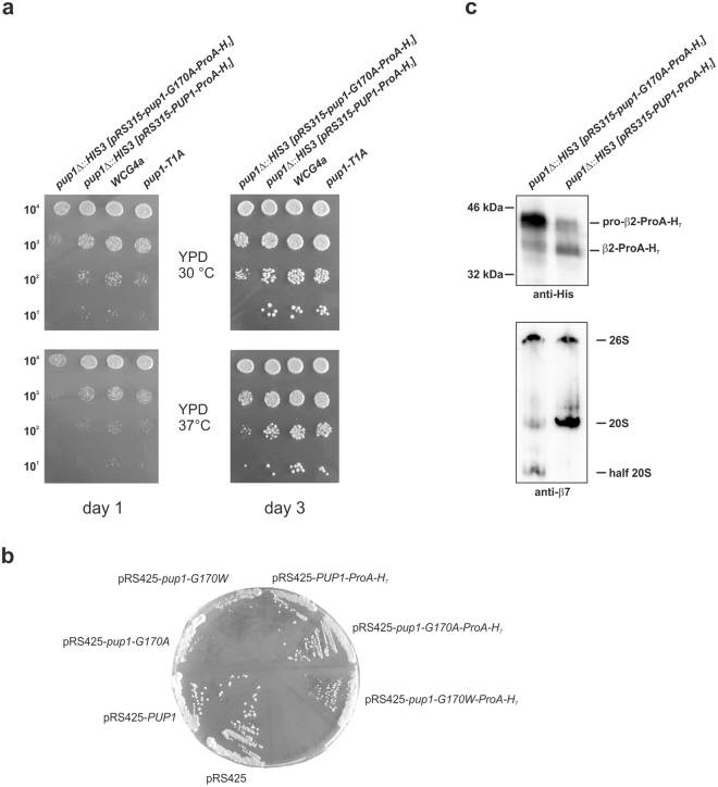
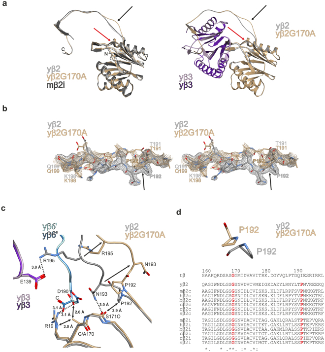
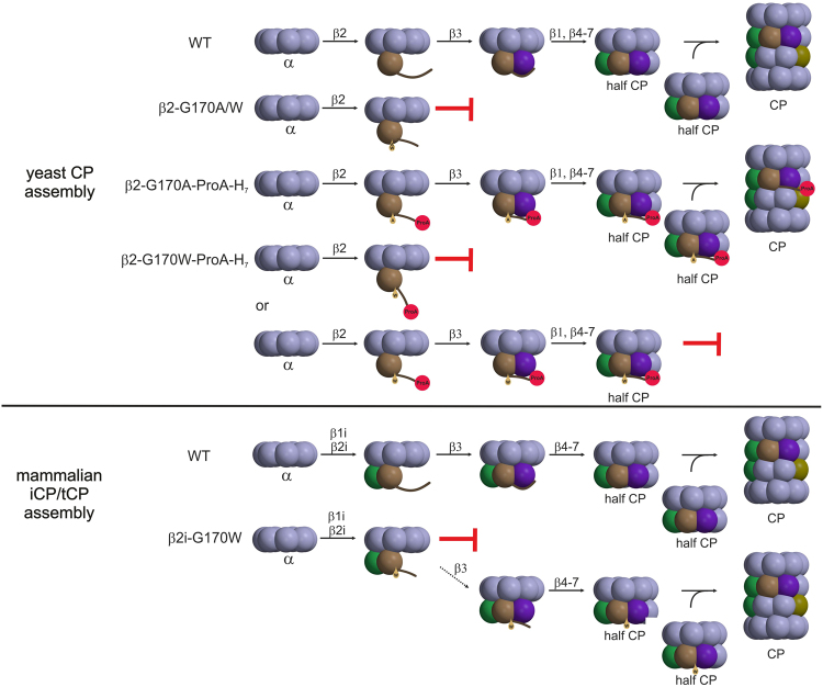
By N-ethyl-N-nitrosourea (ENU) mutagenesis, we generated the mutant mouse line TUB6 that is characterised by severe combined immunodeficiency (SCID) and systemic sterile autoinflammation in homozygotes, and a selective T cell defect in heterozygotes. The causative missense point mutation results in the single amino acid exchange G170W in multicatalytic endopeptidase complex subunit-1 (MECL-1), the β2i-subunit of the immuno- and thymoproteasome. Yeast mutagenesis and crystallographic data suggest that the severe TUB6-phenotype compared to the MECL-1 knockout mouse is caused by structural changes in the C-terminal appendage of β2i that prevent the biogenesis of immuno- and thymoproteasomes. Proteasomes are essential for cell survival, and defective proteasome assembly causes selective death of cells expressing the mutant MECL-1, leading to the severe immunological phenotype. In contrast to the immunosubunits β1i (LMP2) and β5i (LMP7), mutations in the gene encoding MECL-1 have not yet been assigned to human disorders. The TUB6 mutant mouse line exemplifies the involvement of MECL-1 in immunopathogenesis and provides the first mouse model for primary immuno- and thymoproteasome-associated immunodeficiency that may also be relevant in humans.


Conflict of interest statement

The authors declare no competing interests.

Figures

Figure 1
Figure 1
Phenotype of heterozygous TUB6 mutants characterised by numerical and functional T cell defect. (a) T cell frequencies in peripheral blood from heterozygous mutants and littermate controls shown as box plots (pooled data from different experiments, WT n = 40, het n = 71) and representative dot plots (upper panel, gated on living CD45+ cells), and CD4 versus CD8 profiles of T cells (lower panel, gated on living CD45+ CD3+ TCRβ+ cells). (bf) Challenge of heterozygous TUB6 mutants with a low dose of 5000 colony forming units (CFU) (b) or 1000 CFU (cf) Listeria. (b,d) Bacterial loads in indicated organs from infected mice on day 3 (b) and day 7 (d) p.i. Points indicate individual mice. DL detection limit. (c) Survival of infected mice. ****P < 0.0001, Log-rank (Mantel-Cox) test. (e,f) Analysis of antigen-specific T cell response on day 7 p.i. Relative proportions of H-2Kd/LLO91–99 multimer+ cells as percentages of total splenocytes (e) and IFN-γ expressing CD8+ T cells in response to stimulation with dimethyl sulfoxide (DMSO, negative control), peptide LLO91–99 or phorbol 12-myristate 13-acetate (PMA) and ionomycin (f). For flow cytometry dot plots please refer to Supplementary Fig. S1.
Figure 2
Figure 2
Phenotypic comparison of 8-week-old homozygous TUB6 mutants and wild type littermates. (a) Body weight data (n = 8–10 per group). (b) Flow cytometric profile of peripheral blood leukocytes (pooled, hom n = 11, WT n = 44). The right box plot shows granulocyte counts after equal acquisition time per sample, normalised to the mean of wild types. For flow cytometry dot plots see Supplementary Fig. S2. (ce) Representative (immuno-)histological images of spleen (c), neck skin (e) and further indicated organs (d) stained with HE (c,e), anti-CD3 or anti-B220 (c) as indicated, or anti-MPO (d). (f) Cytokine concentration in plasma.
Figure 3
Figure 3
Phenotype of neonatal TUB6 mutants. Flow cytometric analysis of indicated cell populations from 2–5-day-old pups: splenocyte frequencies (a) and CD8SP and CD4SP thymocytes (b). a and b show pooled data from 3 litters analysed on 3 different days (n = 5 WT, 4 het, 4 hom). (c) Analysis of thymic stroma: cortical thymic epithelial cells (cTECs: Epcam+Ly51+) and medullary thymic epithelial cells (mTECs: Epcam+Ly51), gated on CD45 cells (n = 2), shown as percentages of CD45 cells or as total cell counts. (ac) For flow cytometry dot plots see Supplementary Fig. S3.
Figure 4
Figure 4
Underlying genetic alteration identified as missense point mutation in Psmb10 at a highly conserved position. (a) Sanger sequencing of the Psmb10 gene exon 7 shows the G > T nucleotide substitution leading to an amino acid exchange from Gly to Trp in TUB6 mice. (b) Multi-species alignment revealed the strict evolutionary conservation of glycine 170 (indicated by a red arrow). Amino acid numbers are assigned according to the sequence alignment to the β-subunit of Thermoplasma acidophilum.
Figure 5
Figure 5
Overexpression of mutated MECL-1 causes cell death in murine splenocytes. (a) Schematic illustration of the GFP (green fluorescent protein) reporter constructs used for transduction. Fusion of MECL-1 and GFP ensures co-expression of both proteins. To rule out fusion effects, two different linkers between MECL-1 and GFP were used: In the first setting the P2A self-cleavage motif hydrolyses the protein chain, resulting in two separate proteins. In the second setting the self-cleavage ability of P2A was abrogated by a mutation, leading to expression of one fused MECL-1 GFP protein. (b) Kinetics of the GFP signal detected in the splenocyte population after transduction. Plots show changes in GFP signal relative to transduction efficacy. After transduction, T cells were stimulated with anti-CD3 and anti-CD28 antibodies (left panel) or IL-2 and IL-15 (right panel). (c) Apoptotic cells identified by Annexin V staining 36 h after transduction are plotted as percentages of transduced (GFP+) cells. Data from one representative overexpression experiment are shown, in total the experiment was performed twice with similar results. In each experiment splenocytes were transduced with two different virus supernatants. Each point in the dot plots shows data from one separately transduced splenocyte sample.
Figure 6
Figure 6
Yeast mutagenesis experiments. (a) Drop test of mutant yeast strains. Indicated cell numbers were spotted on YPD plates and incubated for 1–3 days either at 30 or 37 °C. Growth of the yeast β2 (pup1) G170A-ProA-H7 mutant is retarded compared to the respective wild type strains and compared to the β2 loss of function mutant T1A. (b) Overexpression of mutant yeast β2-subunits. Wild type yeast (WCG4a) was transformed by either the empty 2 µ plasmid pRS425 or variants thereof encoding wild type PUP1 or mutant pup1 genes. Yeasts were streaked on CM Leu plates (to select for the plasmid pRS425) and grown at 30 °C. (c) Western blot of equal amounts of β2 (pup1) G170A-ProA-H7 and β2 (PUP1)-ProA-H7 cell lysates separated by SDS-PAGE (upper panel) or native PAGE (lower panel). Anti-His immunoblotting of denatured lysates reveals that the mutant accumulates immature β2 (pro-β2), while in wild type cells the processed version of β2 is more abundant. In agreement, probing native lysates with an anti-β7 antibody visualises the accumulation of half 20S proteasomes in the mutant. Note, β7 is the last proteasome subunit that is incorporated into half proteasome precursors. Full-length western blots are presented in Supplementary Fig. S6.
Figure 7
Figure 7
Crystal structure of the yeast β2-G170A mutant proteasome. (a) Ribbon illustration of the G170A mutant yeast proteasome subunit β2 (brown) superimposed onto the wild type counterpart (grey) and the mouse iCP subunit β2i (black) on the left; association of the subunits β2 and β3 (purple) is shown on the right. The site of mutation and the resulting conformational changes in the β2 C-terminal tail are marked by red and black arrows, respectively. (b) Stereo representation of the 2FO-FC omit electron density maps for the G170A mutant (brownish) and wild type yβ2 subunit (grey) contoured to 1σ. Note the structural changes and the increased flexibility of the loop segment in the mutant structure. The flip of Pro192 is marked by a black arrow. (c) Hydrogen-bonding network around the site of mutation. Exchange of Gly170 in subunit yβ2 results in the reorientation of residues 192–196 (black arrows). Hereby hydrogen bonds (black dotted lines) within yβ2 and with the neighbouring subunits yβ3 and yβ6′ (of the opposite half proteasome) are broken. Steric clashes are marked by black double arrows. (d) The strictly conserved Pro192 is supposed to orient the β2 C-terminus for association with the adjacent β3 subunit. In the mutant crystal structure, flipping of Pro192 changes the conformation of the β2 C-terminus. Species abbreviation in sequence alignment: t: T. acidophilum; y: yeast; m: mouse; h: human; b: Bos taurus; c: Canis familiaris; r: Rattus norvegicus; s: Sus scrofa; amino acid numbers are assigned according to the β-subunit of T. acidophilum.
Figure 8
Figure 8
Model of CP assembly and proposed impact of the TUB6 mutation on iCP/tCP biogenesis. The mutations G170W and G170A in the yβ2 proteasome subunit abrogate assembly of the yeast CP (upper panel). Since the β2-G170A-Pro-H7 mutant accumulates half CPs (Fig. 6c), the β2-G170W-Pro-H7 mutant is likely to suffer from a defect concerning half CP dimerisation. Similarly, the TUB6 mutation G170W in the proteasome subunit β2i leads to a major arrest in precursor complex processing and an inefficient, minor progression to mature iCP/tCP (lower panel). Colour code: β1 (green), β2 (brown), β3 (violet), β5 (olive).

References

    1. Hershko A, Ciechanover A, Varshavsky A. Basic medical research award. The ubiquitin system. Nat. Med. 2000;6:1073–1081. doi: 10.1038/80384. - DOI - PubMed
    1. Pamer E, Cresswell P. Mechanisms of MHC class I-restricted antigen processing. Annu. Rev. Immunol. 1998;16:323–358. doi: 10.1146/annurev.immunol.16.1.323. - DOI - PubMed
    1. Huber EM, et al. Immuno- and constitutive proteasome crystal structures reveal differences in substrate and inhibitor specificity. Cell. 2012;148:727–738. doi: 10.1016/j.cell.2011.12.030. - DOI - PubMed
    1. Marques AJ, Palanimurugan R, Matias AC, Ramos PC, Dohmen RJ. Catalytic mechanism and assembly of the proteasome. Chem. Rev. 2009;109:1509–1536. doi: 10.1021/cr8004857. - DOI - PubMed
    1. Groettrup M, Kirk CJ, Basler M. Proteasomes in immune cells: more than peptide producers? Nat. Rev. Immunol. 2010;10:73–78. doi: 10.1038/nri2687. - DOI - PubMed

Publication types

Substances