. 2018 Apr 13;9(1):1444.
doi: 10.1038/s41467-018-03614-0.

A TAD boundary is preserved upon deletion of the CTCF-rich Firre locus

Affiliations

A TAD boundary is preserved upon deletion of the CTCF-rich Firre locus

A Rasim Barutcu et al. Nat Commun. .

Abstract

The binding of the transcriptional regulator CTCF to the genome has been implicated in the formation of topologically associated domains (TADs). However, the general mechanisms of folding the genome into TADs are not fully understood. Here we test the effects of deleting a CTCF-rich locus on TAD boundary formation. Using genome-wide chromosome conformation capture (Hi-C), we focus on one TAD boundary on chromosome X harboring ~ 15 CTCF binding sites and located at the long non-coding RNA (lncRNA) locus Firre. Specifically, this TAD boundary is invariant across evolution, tissues, and temporal dynamics of X-chromosome inactivation. We demonstrate that neither the deletion of this locus nor the ectopic insertion of Firre cDNA or its ectopic expression are sufficient to alter TADs in a sex-specific or allele-specific manner. In contrast, Firre's deletion disrupts the chromatin super-loop formation of the inactive X-chromosome. Collectively, our findings suggest that apart from CTCF binding, additional mechanisms may play roles in establishing TAD boundary formation.


Conflict of interest statement

The authors declare no competing interests.

Figures
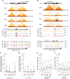
Fig. 1
Fig. 1
Firre is consistently located at a TAD boundary and harbors several CTCF sites. a Human Hi-C heatmaps showing ± 1 Mb of the FIRRE locus in RPE-1 (female), K562 (female), HMEC (male), and NHEK (unisex) cell lines (upper panel), and CTCF ChIP-seq signals across the Firre locus (lower panel). b Mouse Hi-C heatmaps depicting the Firre locus in mESCs (male), CH12 (female), Patski (female), and mouse neuronal stem cells (mNSCs) (unknown sex, upper panel), and a zoom-in of the CTCF ChIP-seq signal (lower panel). c, d Box plot showing the number of CTCF peaks for each sliding window on the (c) human and (d) mouse chromosome X. The bin containing the human and mouse Firre genes is shown with a red dot. e, f Boxplot showing the TAD boundary scores for all the boundaries on the X chromosome in different cell lines in (e) human and in (f) mouse. The TAD boundary that contains Firre is shown with a red dot. Error bars: s.d.
Fig. 2
Fig. 2
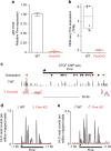
Validation of Firre knockout in MEFs. a qRT-PCR analysis of Firre expression in wild-type and knockout MEFs. Error bars: s.e.m. b Plot showing transcripts per million (TPM) values for wild-type and Firre KO MEF RNA-seq. Error bars: s.d. c CTCF ChIP-seq signal tracks showing the complete loss of CTCF binding at the Firre locus in Firre KO MEFs (mm9, chrX:47.8–49 Mb). d, e Hi-C reads per million (RPM) values for the Firre locus in (e) wild-type and (f) Firre KO MEFs
Fig. 3
Fig. 3
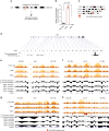
Firre KO does not result in disruption of TAD boundaries. a-d Hi-C heatmaps showing ± 5 Mb of the Firre gene locus (mm9, chr.X: 45–51 Mb) in female wild-type and Firre KO MEFs, (b) male wild-type and Firre KO MEFs, (c) allele-specific haploid chromosomes for female Cast (wild type) and C57BL6 (Firre KO), and (d) male C57BL6 (Firre KO). The TAD boundaries and the insulation plot for each Hi-C dataset is depicted below. e Dot plots showing the boundary strength of the Firre-centered and the neighboring TAD boundaries in wildtype (gray) and Firre KO (red) samples. The TAD insulation scores of all TAD boundaries on chromosome X is shown as boxplots on the right panel. f Hi-C heatmaps from wild-type (grown on 2i) and Firre KO mouse embryonic stem cells (mESCs) (grown on feeders + 2i) showing ± 5 Mb of the Firre locus. g, h Boxplot showing the inter-TAD interaction frequency between the TAD domains neighboring the Firre locus in wild-type (gray) and Firre KO (red) cells (g) in female MEFs and (h) in mESCs. Error bars: s.d. (*p-value: Wilcoxon rank-sum test)
Fig. 4
Fig. 4
TAD boundaries are preserved upon ectopic Firre cDNA insertion and its induced expression at target sites. a Cartoon depicting the approach for the generation of the transgenic MEFs with endogeous Firre KO and ectopic Firre cDNA insertions. b qRT-PCR showing the induced expression of the Firre cDNA in wild type, DOX, and DOX+ conditions. Error bars: s.e.m. (*p-value: t-test). c Chromosome ideograms showing the Firre cDNA insertion sites on four different chromosomes. d CTCF ChIP-seq signal from DOX transgenic MEFs for each of the exons of Firre cDNA at randomly inserted loci. As the transgenic MEFs harbor an endogenous Firre deletion, the intronic regions did not harbor any ChIP-seq signal. e-h Hi-C heatmaps showing the TAD organization, TAD boundary position, and the insulation plots for male Firre KO, DOX, and DOX+ samples ± ~ 5 Mb of Firre cDNA insertion sites on (e) chr 8, (f) chr 10, (g) chr 11, and (h) chr 15
Fig. 5
Fig. 5
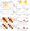
Firre KO results in a loss of Firre-DXZ4, and changes in super-loop interactions in female MEFs. a Hi-C heatmaps at 40 kb resolution in male and female wildtype and Firre KO MEFs showing the interaction frequency between Firre and DXZ4 ± 1 Mb. b Forty-two windows of 40 kb bins were slid across the entire heatmap (2 Mb × 2 Mb in size) to compare the interaction frequency at each position of female and male WT versus KO conditions to the Firre-DXZ4 interaction by t-tests. The p-value distribution in the female or male wild-type samples indicate a drastically enriched significance of Firre-DXZ4 interactions when compared with either the female Firre KO or male samples (*p < 0.05, one-way ANOVA). c Hi-C heatmap at 100 kb resolution showing the zoomed-in interactions between the mouse Firre, DXZ4, x75, and ICCE regions ± 100 kb in female and male wild-type and Firre KO MEFs. d Boxplots showing the interactions among the super-loop regions in wild-type (gray) and Firre KO (red) female (top) and male (bottom) MEF samples. The sample sizes of the boxplots are n = 12 for Firre-DXZ4, DXZ4-x75, and DXZ4-ICCE interactions, and n = 9 for all other combinations. p-value: t-test. Error bars: s.d
Fig. 6
Fig. 6
CRISPR live-cell imaging and 3C validates the changes in super-loop interactions. a Four-color CRISPR live-cell imaging (CLING) from female wild-type and Firre KO MEFs. Firre (red), DXZ4 (green), and x75 (white) loci were simultaneously visualized with Hoechst staining (blue). Pseudo-coloring was used for visual simplicity. Scale bar: 5 μm. b Quantification of the colocalization percentages between Firre-DXZ4, DXZ4-x75, and Firre-x75 between wild-type (black) and Firre KO (red) MEFs. (*p-value: χ2-test, n > 80 nuclei). Error bars: s.e.m. c Chromosome conformation capture (3C) analysis showing the interaction frequency ratios of Firre-DXZ4, DXZ4-x75, and Firre-x75 in female Firre KO vs. wild-type MEFs (*p-value: t-test, n = 3). The black arc indicates 3C enrichment in the wild-type samples, whereas the red arc represents enrichment in the Firre KO samples

Similar articles

Cited by

References

    1. Nora EP, et al. Spatial partitioning of the regulatory landscape of the X-inactivation centre. Nature. 2012;485:381–385. doi: 10.1038/nature11049. - DOI - PMC - PubMed
    1. Dixon JR, et al. Topological domains in mammalian genomes identified by analysis of chromatin interactions. Nature. 2012;485:376–380. doi: 10.1038/nature11082. - DOI - PMC - PubMed
    1. Grubert F, et al. Genetic control of chromatin states in humans involves local and distal chromosomal interactions. Cell. 2015;162:1051–1065. doi: 10.1016/j.cell.2015.07.048. - DOI - PMC - PubMed
    1. Pope BD, et al. Topologically associating domains are stable units of replication-timing regulation. Nature. 2014;515:402–405. doi: 10.1038/nature13986. - DOI - PMC - PubMed
    1. Norton HK, Phillips-Cremins JE. Crossed wires: 3D genome misfolding in human disease. J. Cell. Biol. 2017;216:3441–3452. doi: 10.1083/jcb.201611001. - DOI - PMC - PubMed

Publication types

MeSH terms