. 2018 Apr 13;8(1):5965.
doi: 10.1038/s41598-018-24502-z.

On the use of Parylene C polymer as substrate for peripheral nerve electrodes

Affiliations

On the use of Parylene C polymer as substrate for peripheral nerve electrodes

Natàlia de la Oliva et al. Sci Rep. .

Abstract

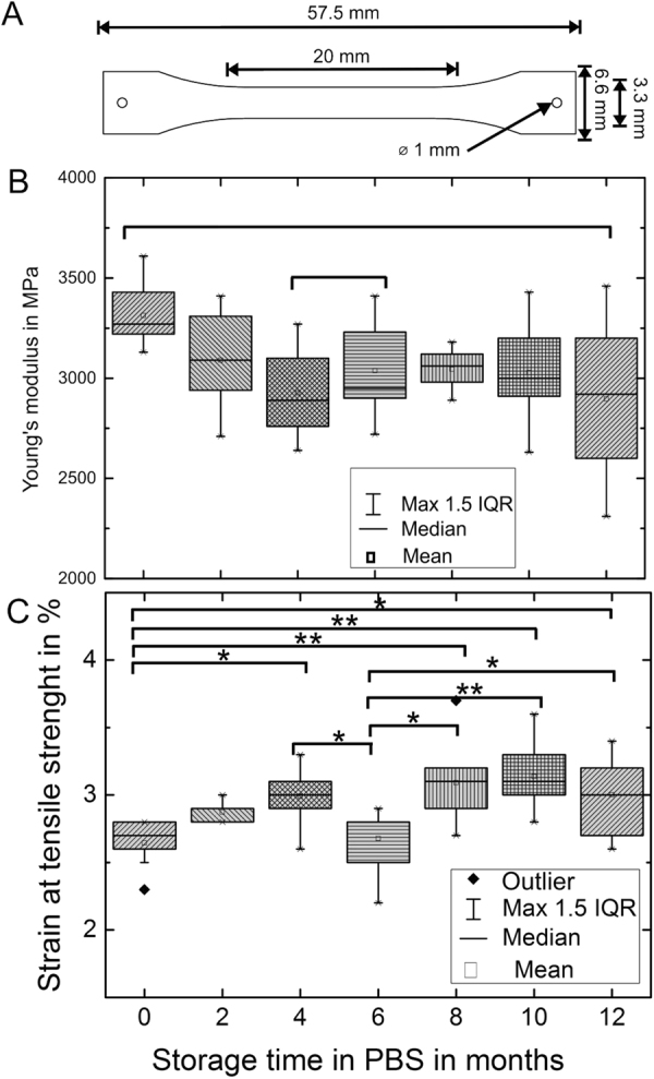
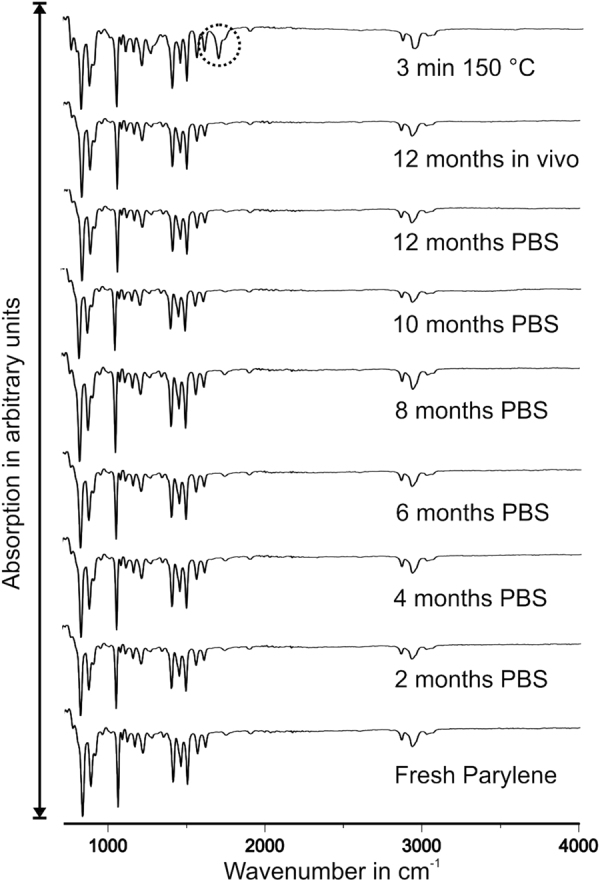
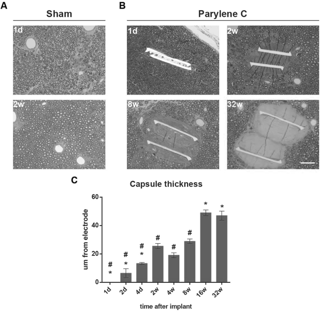
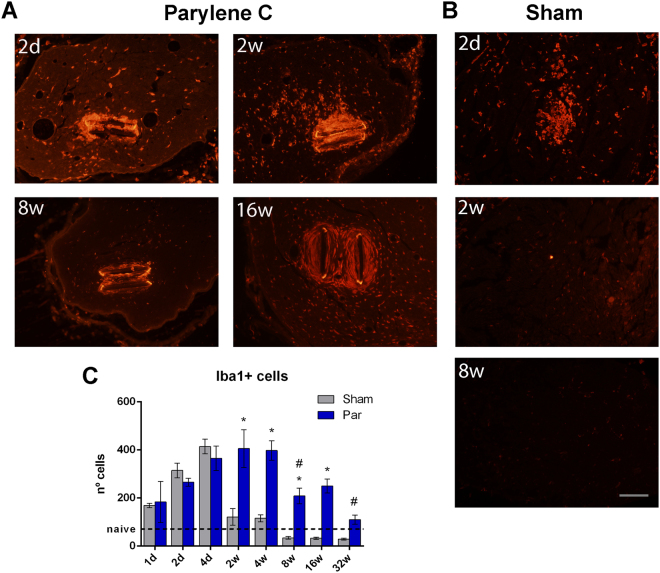
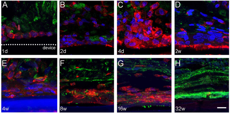
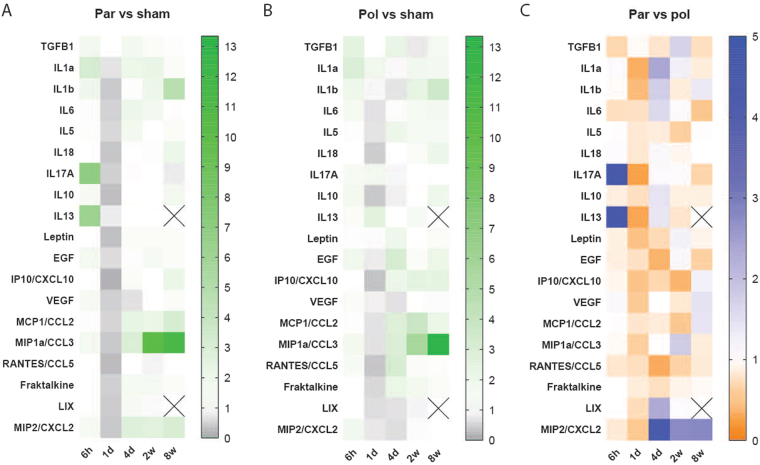
Parylene C is a highly flexible polymer used in several biomedical implants. Since previous studies have reported valuable biocompatible and manufacturing characteristics for brain and intraneural implants, we tested its suitability as a substrate for peripheral nerve electrodes. We evaluated 1-year-aged in vitro samples, where no chemical differences were observed and only a slight deviation on Young's modulus was found. The foreign body reaction (FBR) to longitudinal Parylene C devices implanted in the rat sciatic nerve for 8 months was characterized. After 2 weeks, a capsule was formed around the device, which continued increasing up to 16 and 32 weeks. Histological analyses revealed two cell types implicated in the FBR: macrophages, in contact with the device, and fibroblasts, localized in the outermost zone after 8 weeks. Molecular analysis of implanted nerves comparing Parylene C and polyimide devices revealed a peak of inflammatory cytokines after 1 day of implant, returning to low levels thereafter. Only an increase of CCL2 and CCL3 was found at chronic time-points for both materials. Although no molecular differences in the FBR to both polymers were found, the thick tissue capsule formed around Parylene C puts some concern on its use as a scaffold for intraneural electrodes.


Conflict of interest statement

The authors declare no competing interests.

Figures

Figure 1
Figure 1
(A) Tensile testing samples, according to DIN EN ISO 527-3. (B) Young’s modulus of aged Parylene C samples as box plots. Depiction of 10 samples per measurement set. Maximum length of whiskers is 1.5 times the interquartile range (IQR). ANOVA: F(6,60) = 3.04, p-value = 0.01. Significance (Tukey): *p < 0.05. (C) Strain at tensile strength of aged Parylene C samples as box plots. Depiction of 10 samples per measurement set. Maximum length of whiskers is 1.5 times IQR ANOVA: F(6,60) = 7.39, p-value = 6.1*10−6. Significance (Tukey): *p < 0.05, **p < 0.001.
Figure 2
Figure 2
FTIR spectra of fresh, aged, explanted and intentionally oxidized Parylene C samples. The absorption is presented in arbitrary units. Only oxidized Parylene C exhibits an additional peak.
Figure 3
Figure 3
Progression of tissue capsule formation around the Parylene C device implanted in the nerve. Representative light microscopy images of (A) sham nerves after 1 day and 2 weeks in which no tissue deposition can be appreciated, (B) Implanted nerves with progressive tissue capsule around the device at 1 day, 2, 8 and 32 weeks. (C) Increase in tissue capsule thickness along time. Scale bar = 50 µm. *p < 0.05 vs 2w. #p < 0.05 vs 16w.
Figure 4
Figure 4
Detailed evaluation of tissue capsule by TEM. (AF) Representative images of the tissue capsule at 1 and 2 days, 2, 8, 16 and 32 weeks postimplant. Scale bar = 10 µm. At (A) 1 day, (B) 2 days and (C) 2 weeks only amoeboid cells (arrows) were seen in the vicinity of the device. From (D) week 8 two different zones can be distinguished in the capsule (dotted line), with spindle-shaped cells localized at the periphery of the capsule (arrowheads). After (E) 16 and (F) 32 weeks post-implant, the capsule was mainly formed by spindle-shaped cells and collagen fibers (insets, G–L). Insets show the progressive change from (GH) amoeboid cells at early time points to (IL) spindle-shaped cells, together with an increase in (KL) collagen deposition from 8 weeks. Scale bar = 2 µm.
Figure 5
Figure 5
Iba1 labeling of infiltrating macrophages in the tibial nerve. Representative images of (A) implanted and (B) sham nerves at different time-points. Scale bar = 100 µm. (C) Number of iba1 positive cells in the whole tibial nerve along time. *p < 0.05 vs sham animals. #p < 0.05 vs 2w implanted.
Figure 6
Figure 6
Cellular composition of the capsule. Representative confocal microscopy images labeling against Iba1 and CD90 markers. Only iba1+ macrophages were observed from (A) 1 day to (E) 4 weeks post-implant. From (E) 8 to (F) 16 weeks, CD90+ fibroblasts were located in the periphery of the capsule and by (G) week 32w, CD90+ fibroblasts occupied most of the area of the capsule. Iba1 in red, CD90 in green, DAPI in blue. Scale bar = 5 µm.
Figure 7
Figure 7
Cytokines levels obtained by multiplex analysis. Heatmaps showing the changes in several proteins at different time points in (A) Parylene C and (B) polyimide implanted nerves versus sham nerves. Results are expressed as the mean of the ratio between each group and sham values. (C) Heatmap representing the changes in Parylene C implanted nerves with respect to polyimide implanted nerves. Results expressed as the mean of the ratio between Parylene C and polyimide values. Crossed out squares mean no protein detected.

Similar articles

Cited by

References

    1. Navarro X, et al. A critical review of interfaces with the peripheral nervous system for the control of neuroprostheses and hybrid bionic systems. J. Peripher. Nerv. Syst. 2005;10:229–58. doi: 10.1111/j.1085-9489.2005.10303.x. - DOI - PubMed
    1. Tyler, D. J., Polasek, K. H. & Schiefer, M. A. Peripheral Nerve Interfaces. Nerves and Nerve Injuries2 (Elsevier Ltd., 2015).
    1. del Valle J, Navarro X. Interfaces with the peripheral nerve for the control of neuroprostheses. Int. Rev. Neurobiol. 2013;109:63–83. doi: 10.1016/B978-0-12-420045-6.00002-X. - DOI - PubMed
    1. Badia J, et al. Comparative analysis of transverse intrafascicular multichannel, longitudinal intrafascicular and multipolar cuff electrodes for the selective stimulation of nerve fascicles. J. Neural Eng. 2011;8:36023. doi: 10.1088/1741-2560/8/3/036023. - DOI - PubMed
    1. Oddo CM, et al. Intraneural stimulation elicits discrimination of textural features by artificial fingertip in intact and amputee humans. Elife. 2016;5:1–27. doi: 10.7554/eLife.09148. - DOI - PMC - PubMed

Publication types

MeSH terms