Review
. 2018 Oct;33(4):297-312.
doi: 10.1007/s12928-018-0523-z. Epub 2018 Apr 13.

Contemporary critical limb ischemia: Asian multidisciplinary consensus statement on the collaboration between endovascular therapy and wound care

Affiliations
Review

Contemporary critical limb ischemia: Asian multidisciplinary consensus statement on the collaboration between endovascular therapy and wound care

Osami Kawarada et al. Cardiovasc Interv Ther. 2018 Oct.

Abstract

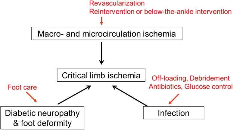
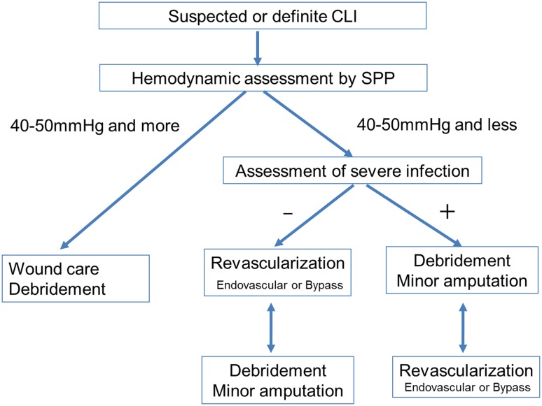
The burden of peripheral artery disease (PAD) and diabetes in Asia is projected to increase. Asia also has the highest incidence and prevalence of end-stage renal disease (ESRD) in the world. Therefore, most Asian patients with PAD might have diabetic PAD or ESRD-related PAD. Given these pandemic conditions, critical limb ischemia (CLI) with diabetes or ESRD, the most advanced and challenging subset of PAD, is an emerging public health issue in Asian countries. Given that diabetic and ESRD-related CLI have complex pathophysiology that involve arterial insufficiency, bacterial infection, neuropathy, and foot deformity, a coordinated approach that involves endovascular therapy and wound care is vital. Recently, there is increasing interaction among cardiologists, vascular surgeons, radiologists, orthopedic surgeons, and plastic surgeons beyond specialty and country boundaries in Asia. This article is intended to share practical Asian multidisciplinary consensus statement on the collaboration between endovascular therapy and wound care for CLI.

Keywords: Bacterial infection; Collaboration; Foot deformity; Interdisciplinary; Ischemia; Peripheral artery disease.


Conflict of interest statement

Osami Kawarada reports honorarium for lectures and advisory board fees from Boston Scientific Corporation, honorarium for lectures and research grants from Terumo, and a consultancy fee from Medtronic. Hsuan-Li Huang reports honorarium for lectures from Boston Scientific and Medtronic. Testuya Nakama reports honorarium for lectures from Abbott Vascular, Boston Scientific, and Medtronic and consulting fee from Boston Scientific and Century Medical Inc. Naoki Fujimura reports consulting fee from W.L. Gore and Associates. Bryan Ping-Yen Yan reports honorarium for lectures from Boston Scientific Corporation, Cook Medical, and Medtronic, research grants from Medtronic and Boston Scientific Corporation, and consultancy fee from Medtronic and Cook Medical. Robbie K George reports honorariums for/consultancy agreements with Medtronic Corporation, Bard Corporation, Cook Medical, and Abbot Vascular. Kan Zen, Shinobu Aayabe, Donghoon Choi, Su Hong Kim, Jiyoun Kim, Taku Kato, Yoshinori Tsubakimoto, Shigeo Ichihashi, Akihiro Higashimori, Masahiko Fujihara, Tomoyasu Sato, Skyi Yin-Chun Pang, Chumpol Wongwanit, Yew Pung Leong, Benjamin Chua, Yoshiaki Yokoi, Hisashi Motomura, and Hideaki Obara report no conflict of interest.

Figures

Fig. 1
Fig. 1
Representative ischemic tissue loss. a Traditional CLI. Traditional CLI such as nondiabetic or non ESRD-related CLI can typically cause dry tissue loss. b Diabetic CLI or ESRD-related CLI. Most of the diabetic or ESRD-related CLI cause wet tissue loss, suggesting significant bacterial infection. This is the most challenging subset of CLI. c Diabetic foot. Although wound appearance is similar to CLI, foot microcirculation is preserved
Fig. 2
Fig. 2
Assessment of the macrocirculation and microcirculation. a Relationship between ABI and probability of ischemic wound healing (modified from reference [15]). In patients without diabetes or renal failure, there is a significant relationship between ABI and wound healing. However, in patients with diabetes or renal failure, which commonly co-exist with critical limb ischemia, no relationship between ABI and wound healing is observed, suggesting that assessing the role of the macrocirculation is limited in the setting of critical limb ischemia. DM diabetes mellitus, CRF chronic renal failure. b Relationship between SPP at the proximal margin of the wound and probability of ischemic wound healing (modified from reference [19]). There is a significant relationship between SPP and wound healing, suggesting that assessment of the microcirculation is important. If SPP is 40–50 mmHg or more, the probability of wound healing is over 90%. Therefore, the SPP cutoff point of the diagnosis of CLI is considered to be 40–50 mmHg and the aim of revascularization is to achieve SPP of 40 mmHg or more
Fig. 3
Fig. 3
Diagnostic algorithm for atherosclerotic CLI, nonatherosclerotic CLI, and conditions other than CLI in patients with rest pain or tissue loss (modified from reference [28])
Fig. 4
Fig. 4
Venous stasis ulcer complicated by peripheral artery disease. a A 74-year-old female presenting with ulcer above the ankle. b Enhanced CT revealed long occlusion in the femoropopliteal artery (arrows). However, SPP around the ankle was almost 40 mmHg, suggesting preservation of microcirculation. c Wound management including compressive bandage facilitated complete wound healing 6 months later without revascularization
Fig. 5
Fig. 5
Complex pathology in critical limb ischemia. CLI can be characterized by multiple disorders. A comprehensive approach tailored to each patient including revascularization, infection control, and foot care is needed
Fig. 6
Fig. 6
Potential for underlying severe infectious disease in patients with critical limb ischemia. a Osteomyelitis. Only a tiny necrotic lesion was visible on the top of the great toe. However, radiography demonstrated osteolysis of the distal phalanx, suggesting osteomyelitis. b Gas gangrene. Extensive gangrene on the dorsum of the foot was visible. Physical examination revealed snowball crepitus and radiography demonstrated the presence of air in the foot, suggesting gas gangrene. c Necrotizing fasciitis. Gangrene on the lateral side of foot with relative sparing of the skin was visible. Given the rapidly progressive course, severe inflammation, and high CRP value, necrotizing fasciitis was suspected. Emergent surgical incision demonstrated extensive necrosis of the subcutaneous fat and fascia with abscess formation
Fig. 7
Fig. 7
Comprehensive strategy for suspected or definite CLI. If the microcirculation is preserved (i.e., SPP is 40–50 mmHg and more), the foot is not affected by critical ischemia. Nonischemic reasons should be investigated. Wound care is the cornerstone of treatment. If the microcirculation is inadequate (i.e., SPP is 40–50 mmHg or less) and no severe bacterial infection is present, revascularization needs to be considered first. If the microcirculation is inadequate (i.e., SPP is 40–50 mmHg or less) and severe bacterial infection is present, debridement should be considered before revascularization
Fig. 8
Fig. 8
Practical collaboration of endovascular therapy and debridement/minor amputation. Caution needs to be undertaken about the development of bacterial infection after revascularization or preexisting severe bacterial infection before revascularization. The wound healing process after revascularization consists of 3 stages such as bleeding and inflammation stage, granulation stage, and epithelialization and reconstitution stage. a A case of development of bacterial infection after revascularization. A 65-year-old man with diabetes, ESRD, and second toe necrosis. Following the first intervention, severe cellulitis and abscess formation developed rapidly and extensively on the plantar aspect of the foot just below the gangrene on the second toe. Note the redness and swelling of the central foot just below the second toe gangrene. Urgent debridement and open drainage was implemented to prevent the further development of sepsis. Subsequently, granulation became evident after clinically driven reintervention. Finally, complete wound healing was achieved 6 months later. b A case of preexisting severe bacterial infection before revascularization. A 58-year-old man with diabetes, ESRD, and extensive infectious gangrene. Note the redness and swelling of the medial foot just below the first toe gangrene. Immediately after the first debridement of infectious tissue to prevent the development of sepsis, the first infrapopliteal intervention was performed. During the process of wound healing, clinically driven reintervention was required twice on the basis of skin perfusion pressure guidance, and a total of three times debridement or minor amputation was also executed. Finally, complete wound healing and gait acquisition were achieved 9 months later (modified from reference [4])
Fig. 9
Fig. 9
Endovascular strategy for multisegment disease or isolated infrapopliteal disease in CLI
Fig. 10
Fig. 10
Representative case of staged procedures for multisegment disease presenting with tissue loss. a A 71-year-old male presenting with unhealing ulcer on the right second toe. Foot SPP was not measurable because of serious foot pain. b Enhanced CT revealed multisegment disease including occlusions in the right extrenal iliac (thin arrow) and femoropopliteal arteries (thick arrows). c Enhanced CT also revealed extensive occlusions in the right anterior tibial and peroneal arteries. d In the first session, right external iliac artery occlusion was successfully recanalized with stents. Given the concomitant femoropopliteal lesion complexity, contrast volume used, and procedure time, long occlusion in the femoropopliteal artery remained untreated in the session. Although rest pain significantly improved, the wound did not significantly heal with the foot SPP only 25 mmHg on the dorsum and 15 mmHg on the plantar, suggesting the need for further intervention. e In the second session, long occlusion in the right femoropopliteal artery was successfully recanalized with stents. However, given the potential of balloon-induced early reocclusion in the crural artery, the concomitant infrapopliteal artery remained untreated. f Foot SPP increased to 77 mmHg on the dorsum and 70 mmHg on the plantar, and the wound completely healed 5 months later
Fig. 11
Fig. 11
One straight-line strategy. a Baseline angiography. b Final angiography. In the setting of multivessel occlusion, the anterior tibial artery was successfully recanalized based on the interpretation of angiographic findings, suggesting the establishment of one straight-line flow to the foot (cited from reference [9])
Fig. 12
Fig. 12
As many as possible strategy. a Baseline angiography. Note triple vessel disease in the infrapopliteal artery. b Post first intervention. During the first intervention, multiple stenotic areas in the peroneal artery were treated because it was technically straightforward. c Post second intervention. Since foot microcirculation after the first intervention was insufficient for wound healing (SPP 15 → 30 mmHg on the dorsum, 13 → 15 mmHg on the plantar), a second intervention was attempted. Since the reconstituted vessel in the distal anterior tibial artery became clear, recanalization of the long occlusion in the anterior tibial artery was successful. SPP increased to 52 mmHg on the dorsum and 42 mmHg on the plantar, and the wound healed completely
Fig. 13
Fig. 13
Schematic representation of foot perfusion in theory and the practical clinical perfusion territory of a single-source artery (modified from reference [4]). a Perfusion in the normal subject. b Clinical perfusion by a single-source artery. In cases of posterior tibial artery occlusion, the practical territory of the anterior tibial artery extends to encompass the normal territory of the adjacent source artery (posterior tibial artery) through the pedal arch, branches, and arterial connections. c A non-healing ulcer on the second toe (plantar side) in the setting of subsequent stenosis in the anterior tibial artery. Endovascular revascularization can be expected to significantly increase overall blood flow to the foot and the wound
Fig. 14
Fig. 14
Types of below-the-ankle lesions. a Separate lesion type. At baseline, the anterior tibial artery lesions (thin arrows) were distinct from the below-the-ankle lesion, which involved an occluded dorsalis pedis artery through the reconstituted segment just above the ankle (thick arrow). Only above-the-ankle intervention for the anterior tibial artery was attempted to establish straight-line blood flow to the foot (cited from reference [61]). Although the occluded dorsalis pedis artery was left untreated, foot skin perfusion pressure increased from 25/32 to 37/64 mmHg (dorsal/plantar), leading to complete wound healing with debridement. b Serial lesion type. Baseline angiography showing extensive serial lesions from the anterior tibial artery to the dorsalis pedis artery beyond the ankle joint (arrow). Below-the-ankle intervention for the occluded dorsalis pedis artery was performed in the single setting with above-the-ankle intervention for the occluded anterior tibial artery to establish straight-line blood flow to the foot. c Isolated lesion type. Baseline angiography showed short segment occlusion in the dorsalis pedis artery (arrow) with diffuse involvement of the peroneal artery and a long occlusion in the posterior tibial to plantar arteries. Below-the-ankle intervention for the occluded dorsalis pedis artery was performed to establish straight-line blood flow to the foot (cited from reference [57])
Fig. 15
Fig. 15
Endovascular strategy for below-the-ankle lesions. In cases where the below-the-ankle lesions are separate from the above-the-ankle lesions (separate lesion type), below-the-ankle intervention is not recommended during the first intervention because complications related to endovascular procedure and post-intervention reocclusion can potentially be catastrophic. In cases of extensive serial lesions beyond the ankle joint (serial lesion type) or isolated below-the-ankle lesions (isolated lesion type), treating the below-the-ankle lesions is recommended in the first intervention to improve foot microcirculation

Similar articles

Cited by

References

    1. Fowkes FG, Rudan D, Rudan I, et al. Comparison of global estimates of prevalence and risk factors for peripheral artery disease in 2000 and 2010: a systematic review and analysis. Lancet. 2013;382:1329–1340. doi: 10.1016/S0140-6736(13)61249-0. - DOI - PubMed
    1. Ogurtsova K, da Rocha Fernandes JD, Huang Y, et al. IDF Diabetes Atlas: global estimates for the prevalence of diabetes for 2015 and 2040. Diabetes Res Clin Pract. 2017;128:40–50. doi: 10.1016/j.diabres.2017.03.024. - DOI - PubMed
    1. Prasad N, Jha V. Hemodialysis in Asia. Kidney Dis (Basel). 2015;1:165–177. doi: 10.1159/000441816. - DOI - PMC - PubMed
    1. Kawarada O, Yasuda S, Huang J, et al. Contemporary infrapopliteal intervention for limb salvage and wound healing: harmonization of revascularization and wound management. Circ J. 2014;78:1540–1549. doi: 10.1253/circj.CJ-14-0060. - DOI - PubMed
    1. Beckman JA, Creager MA, Libby P. Diabetes and atherosclerosis: epidemiology, pathophysiology, and management. JAMA. 2002;287:2570–2581. doi: 10.1001/jama.287.19.2570. - DOI - PubMed

MeSH terms