. 2018 Apr;6(7):e13666.
doi: 10.14814/phy2.13666.

Measurement of cations, anions, and acetate in serum, urine, cerebrospinal fluid, and tissue by ion chromatography

Affiliations

Measurement of cations, anions, and acetate in serum, urine, cerebrospinal fluid, and tissue by ion chromatography

Andrew D Chapp et al. Physiol Rep. 2018 Apr.

Abstract

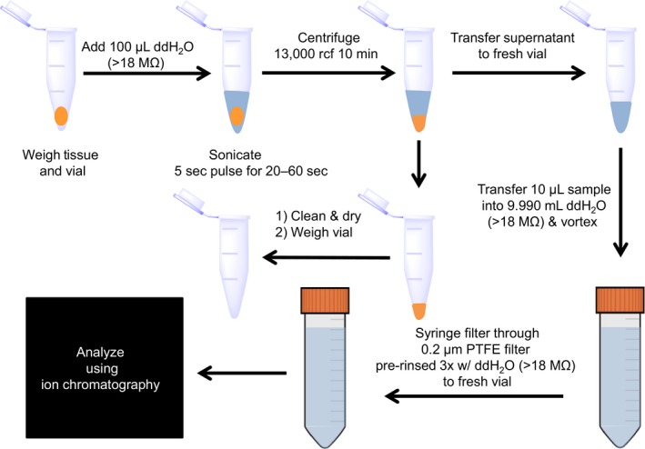
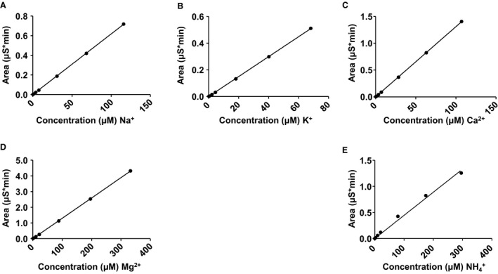
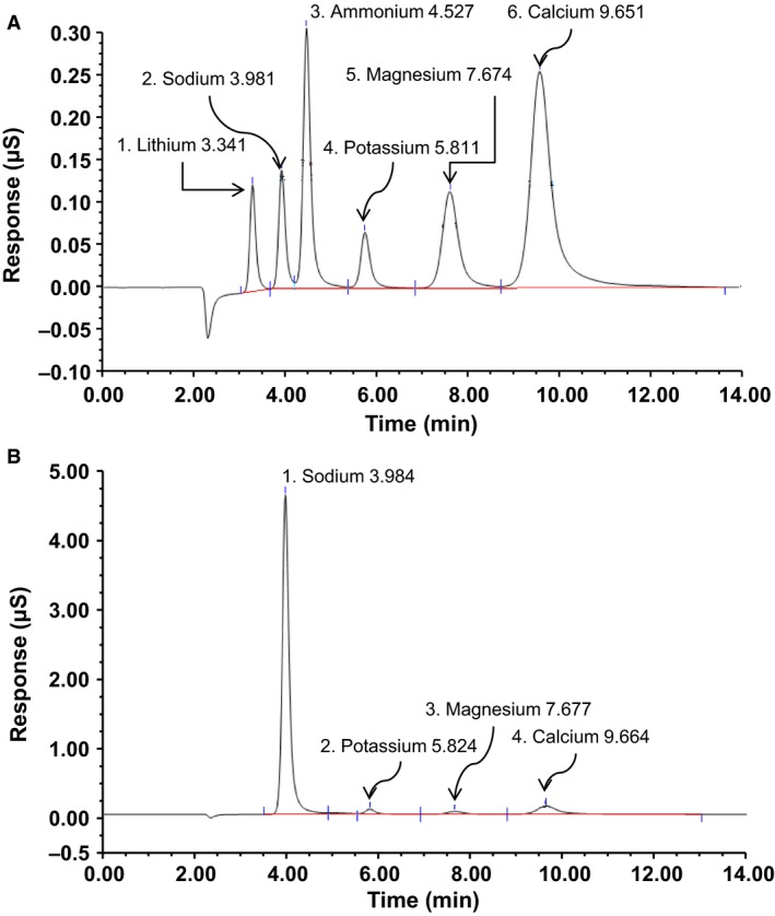
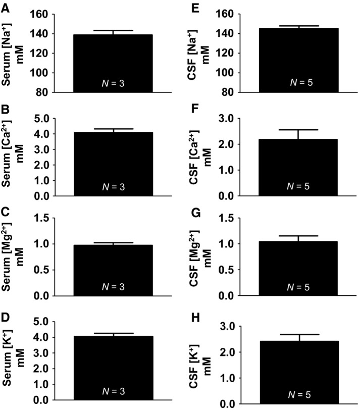
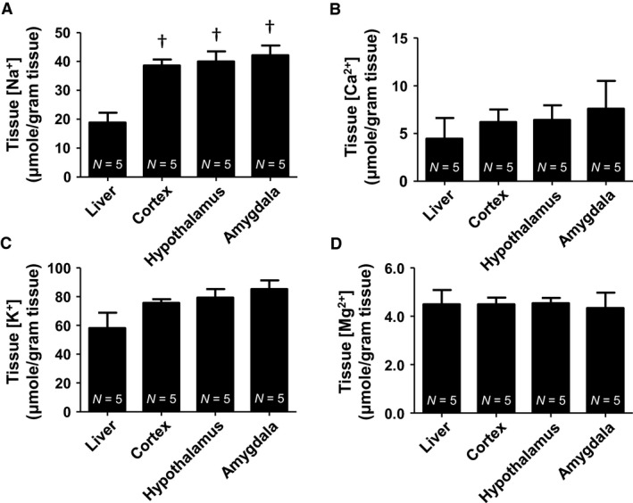
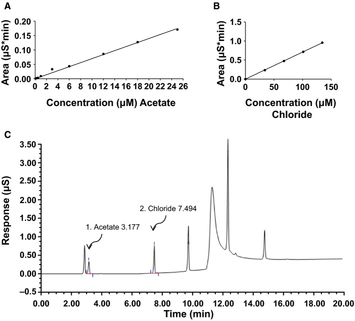
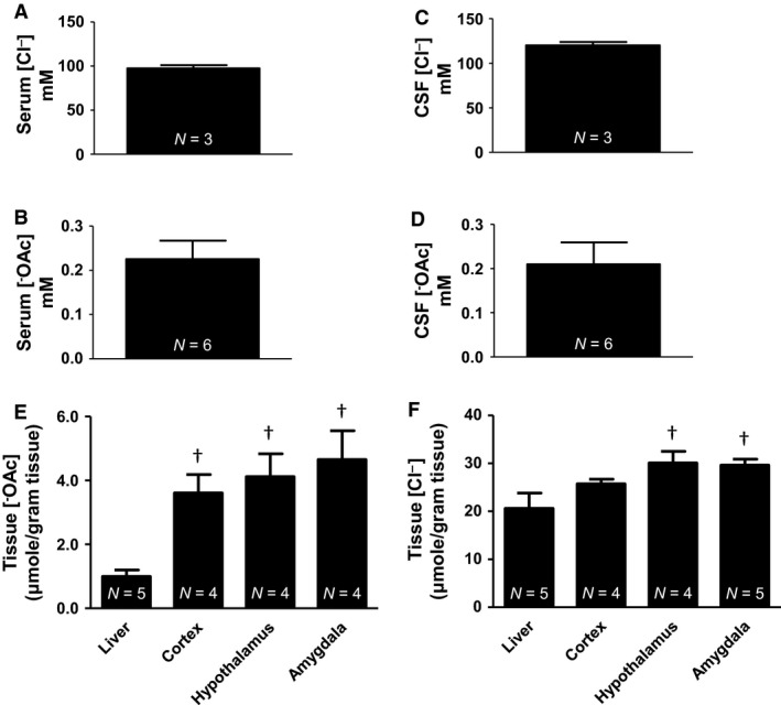
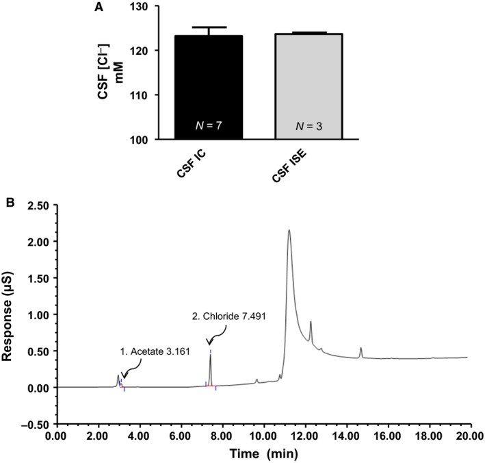
Accurate quantification of cations and anions remains a major diagnostic tool in understanding diseased states. The current technologies used for these analyses are either unable to quantify all ions due to sample size/volume, instrument setup/method, or are only able to measure ion concentrations from one physiological sample (liquid or solid). Herein, we adapted a common analytical chemistry technique, ion chromatography and applied it to measure the concentration of cations; sodium, potassium, calcium, and magnesium (Na+ , K+ , Ca2+ , and Mg2+ ) and anions; chloride, and acetate (Cl- , - OAc) from physiological samples. Specifically, cations and anions were measured in liquid samples: serum, urine, and cerebrospinal fluid, as well as tissue samples: liver, cortex, hypothalamus, and amygdala. Serum concentrations of Na+ , K+ , Ca2+ , Mg2+ , Cl- , and - OAc (mmol/L): 138.8 ± 4.56, 4.05 ± 0.21, 4.07 ± 0.26, 0.98 ± 0.05, 97.7 ± 3.42, and 0.23 ± 0.04, respectively. Cerebrospinal fluid concentrations of Na+ , K+ , Ca2+ , Mg2+ , Cl- , and - OAc (mmol/L): 145.1 ± 2.81, 2.41 ± 0.26, 2.18 ± 0.38, 1.04 ± 0.11, 120.2 ± 3.75, 0.21 ± 0.05, respectively. Tissue Na+ , K+ , Ca2+ , Mg2+ , Cl- , and - OAc were also measured. Validation of the ion chromatography method was established by comparing chloride concentration between ion chromatography with a known method using an ion selective chloride electrode. These results indicate that ion chromatography is a suitable method for the measurement of cations and anions, including acetate from various physiological samples.

Keywords: Anion; cations; electrolyte; ion chromatography.


Figures

Figure 1
Figure 1
Flow diagram for sample preparation. Tissue samples are weighed in their vials, and 100 μL ddH2O (>18 MΩ) added. Samples are sonicated for 20–60 sec in 5 sec pulses at 20 kHz or until well homogenized. The samples are centrifuged at 13,000 rcf for 10 min and the supernatant removed and transferred to a fresh 1.5 mL centrifuge tube. Empty tissue vials are washed, dried, and weighed to determine the mass of tissue. Liquid samples are prepared by adding 10 μL sample with 9.990 mL ddH2O (>18 MΩ), vortexed and syringe filtered into fresh vials. Samples are then run on ion chromatography to determine cation and anion concentrations.
Figure 2
Figure 2
Cation standard curves. (A) Sodium (Na+) standard curve. (B) Potassium (K+) standard curve. (C) Calcium (Ca2+) standard curve. (D) Magnesium (Mg2+) standard curve. (E) Ammonium (NH 4 +) standard curve. A linear regression line was fitted to each standard curve and unknown cation concentrations determined from the slope of the line.
Figure 3
Figure 3
Representative cation chromatograms from IC. (A) Representative cation standards chromatogram containing lithium (Li+), sodium (Na+), ammonium (NH 4 +), potassium (K+), magnesium (Mg2+), and calcium (Ca2+). (B) Representative cation chromatogram from a cerebrospinal fluid (CSF) sample obtained from a Sprague‐Dawley rat.
Figure 4
Figure 4
Cation concentrations from serum and CSF. (A) Serum sodium (138.8 ± 4.56 mmol/L). (B) Serum potassium (4.05 ± 0.21 mmol/L). (C) Serum calcium (4.07 ± 0.26 mmol/L). (D) Serum magnesium (0.98 ± 0.05 mmol/L). (E) CSF sodium (145.1 ± 2.81 mmol/L). (F) CSF potassium (2.41 ± 0.26 mmol/L). (G) CSF calcium (2.18 ± 0.38 mmol/L). (H) CSF magnesium (1.04 ± 0.11 mmol/L).
Figure 5
Figure 5
Cation concentrations from tissue samples. (A) Tissue Na+ (μmol/gram tissue): Liver (18.86 ± 3.42), cortex (38.58 ± 2.09), hypothalamus (39.98 ± 3.54), and amygdala (42.16 ± 3.37). (B) Tissue K+ (μmol/gram tissue): Liver (58.14 ± 10.66), cortex (75.62 ± 2.56), hypothalamus (79.22 ± 5.97), and amygdala (85.21 ± 6.08). (C) Tissue Ca2+ (μmol/gram tissue): Liver (4.45 ± 2.16), cortex (6.20 ± 1.32), hypothalamus (6.41 ± 1.55), and amygdala (7.59 ± 2.90). (D) Tissue Mg2+ (μmol/gram tissue): Liver (4.49 ± 0.59), cortex (4.49 ± 0.28), hypothalamus (4.53 ± 0.22), and amygdala (4.34 ± 0.63). (†vs. control one‐way ANOVA.
Figure 6
Figure 6
Anion standard curves and representative chromatogram. (A) Acetate standard curve. (B) Chloride standard curve. (C) Representative anion standard chromatogram from IC. Anion peaks were well resolved. A linear regression line was fitted to each standard curve and unknown anion concentrations determined from the slope of the line.
Figure 7
Figure 7
Summary data for anions. (A) Serum Cl (97.7 ± 3.42 mmol/L). (B) Serum OAc (0.23 ± 0.04 mmol/L). (C) CSF Cl (120.2 ± 2.81 mmol/L). (D) CSF OAc (0.21 ± 0.05 mmol/L). (E) Tissue OAc (μmol/gram tissue): Liver (1.00 ± 0.19), cortex (3.61 ± 0.58), hypothalamus (4.12 ± 0.72), and amygdala (4.65 ± 0.90). (F) Tissue Cl (μmol/gram tissue): Liver (20.57 ± 3.20), cortex (25.72 ± 0.95), hypothalamus (30.09 ± 2.40), and amygdala (29.61 ± 1.25). (†vs. liver one‐way ANOVA).
Figure 8
Figure 8
Method validation between IC and ISE. (A) Summary data for CSF chloride concentrations measured with IC (123.2 ± 1.94 mmol/L) and ISE (123.7 ± 0.33 mmol/L). There was no statistical difference between CSF chloride concentrations from IC vs. ISE. (B) Representative anion chromatogram from IC for a tissue sample from the cortex of a Sprague‐Dawley rat.
Figure 9
Figure 9
Known ester derivatization reactions with acetate. Reaction 1 shows acetate derivatization to benzyl acetate using benzyldimethylammonium hydroxide as an ester derivatizing agent.(Richards et al. 1975) Reaction 2 shows acetate derivatization to propyl acetate using methylchloroformate as a derivatizing agent, followed by a subsequent reaction with propyl alcohol.(Tumanov et al. 2016) Any amino acids, carboxylic acids or nucleophiles remaining in samples are capable of reacting with derivatizing reagents and forming side products, drastically reducing the apparent concentration of SCFA and or leading to wide ranges for reported values. BDMPH, (benzyldimethylphenylammonium hydroxide; MCF, methylchloroformate.

References

    1. Amtorp, O. , and Sorensen S. C.. 1974. The ontogenetic development of concentration differences for protein and ions between plasma and cerebrospinal fluid in rabbits and rats. J. Physiol. 243:387–400. - PMC - PubMed
    1. Avery, D. H. , Overall J. E., Calil H. M., and Hollister L. E.. 1983. Plasma calcium and phosphate during alcohol intoxication. Alcoholics versus nonalcoholics. J. Stud. Alcohol 44:205–214. - PubMed
    1. Chapp, A. D. , Gui L., Huber M. J., Liu J., Larson R. A., Zhu J., et al. 2014. Sympathoexcitation and pressor responses induced by ethanol in the central nucleus of amygdala involves activation of NMDA receptors in rats. Am. J. Physiol. Heart Circ. Physiol. 307:H701–H709. - PMC - PubMed
    1. Christensen, J. D. , Barrere B. J., Boada F. E., Vevea J. M., and Thulborn K. R.. 1996. Quantitative tissue sodium concentration mapping of normal rat brain. Magn. Reson. Med. 36:83–89. - PubMed
    1. Chung, J.‐W. , Ryu W.‐S., Kim B. J., and Yoon B.‐W.. 2015. Elevated calcium after acute ischemic stroke: association with a poor short‐term outcome and long‐term mortality. J. Stroke 17:54–59. - PMC - PubMed

Publication types

LinkOut - more resources