. 2018 May 22;114(10):2376-2385.
doi: 10.1016/j.bpj.2018.03.008. Epub 2018 Apr 11.

Chromatin Fiber Folding Directed by Cooperative Histone Tail Acetylation and Linker Histone Binding

Affiliations

Chromatin Fiber Folding Directed by Cooperative Histone Tail Acetylation and Linker Histone Binding

Gavin D Bascom et al. Biophys J. .

Abstract

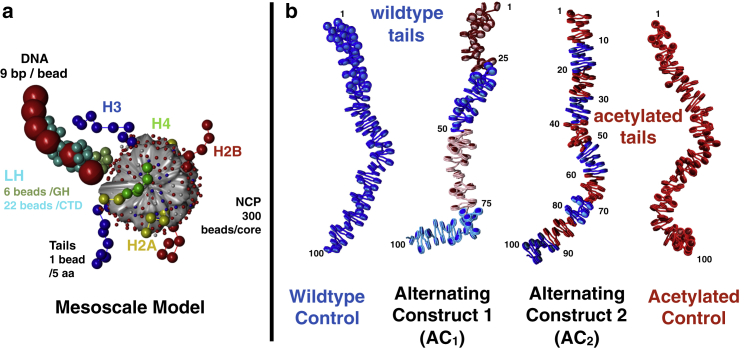
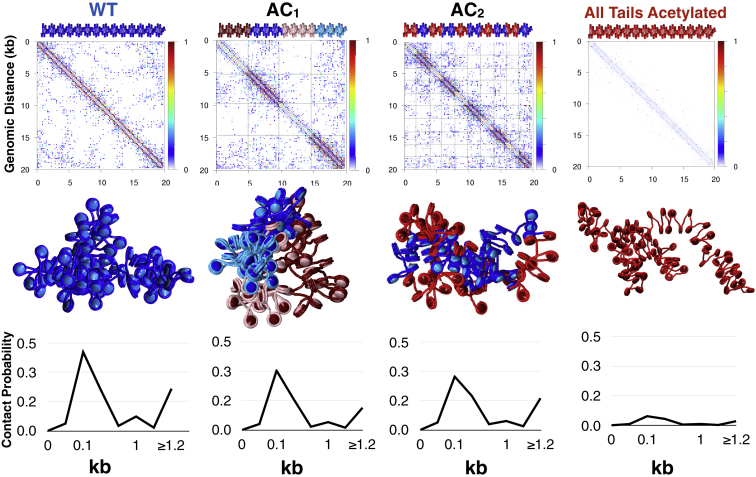
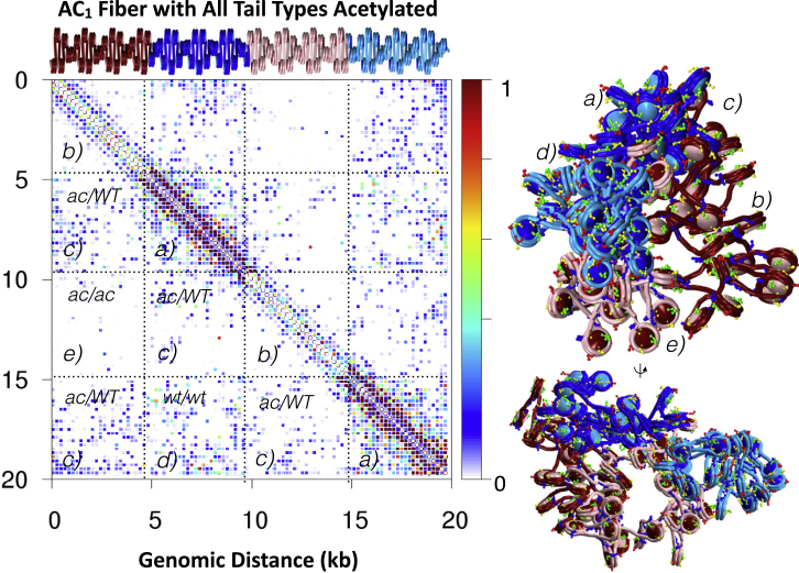
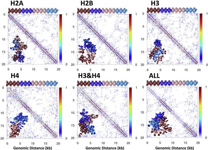
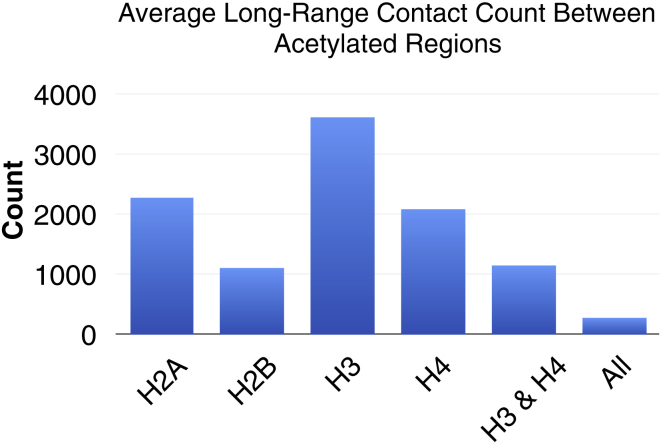
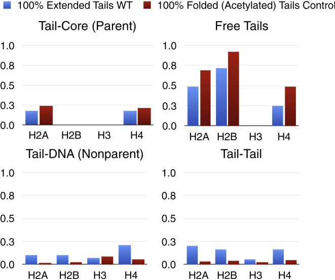
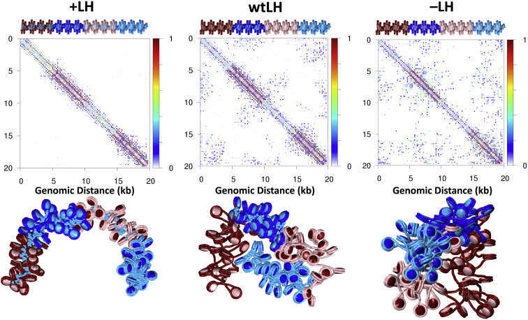
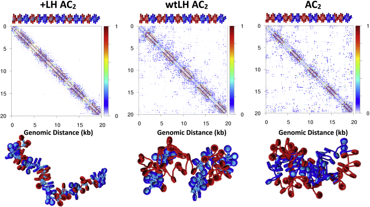
In eukaryotic chromatin, islands of histone tail acetylation are found near transcription start sites and enhancers, whereas linker histones (LHs) are localized in intergenic regions with wild-type (WT) histone tails. However, the structural mechanisms by which acetylation, in combination with LH binding, modulates chromatin compaction and hence transcription regulation are unknown. To explore the folding propensity by which these features may govern gene expression, we analyze 20 kb fibers that contain regularly spaced acetylation islands of two sizes (2 or 5 kb) with various LH levels by mesoscale modeling. Specifically, we investigate the effect of acetylating each histone tail (H3, H4, H2A, and H2B) individually, in combination (H3 and H4, or all tails), and adding LH to WT regions. We find that fibers with acetylated H4 tails lose local contacts (<1 kb) and fibers with all tails acetylated have decreased long-range contacts in those regions. Tail interaction plots show that this opening of the fiber is driven by the loss of tail-tail interactions in favor of tail-parent core interactions and/or increase in free tails. When adding LH to WT regions, the fibers undergo hierarchical looping, enriching long-range contacts between WT and acetylated domains. For reference, adding LH to the entire fiber results in local condensation and loss of overall long-range contacts. These findings highlight the cooperation between histone tail acetylation and regulatory proteins like LH in directing folding and structural heterogeneity of chromatin fibers. The results advance our understanding of chromatin contact domains, which represent a pivotal part of the cell cycle, diseased states, and differentiation states in eukaryotic cells.


Figures

Figure 1
Figure 1
Chromatin model and fiber systems. (a) Our mesoscale model building block, which consists of linker DNA beads, a rigid nucleosome core with discrete charges, flexible histone tail beads, and flexible LH beads. (b) Starting configurations for four chromatin fiber systems studied with varying levels of acetylation are shown. From left to right: wild-type (WT) system, Alternating Construct 1 (AC1) system consisting of alternating WT (blue) and acetylated regions (magenta or pink) with 25 nucleosome segments, Alternating Construct 2 (AC2) with 10 nucleosome segments, and the all-acetylated control (red). Histone tails are not rendered here for clarity. To see this figure in color, go online.
Figure 2
Figure 2
Probability contact maps, fiber renderings, and short-range internucleosome contact probability profiles for WT, AC1, AC2 and acetylated control fibers (see Fig. 1). Regions of acetylated histone tails (which are more tightly folded) are rendered in red or pink, whereas WT regions are colored blue. To see this figure in color, go online.
Figure 3
Figure 3
Structural analysis of AC1 fibers with all tail types acetylated within the noted 25-nucleosome segment. The contact map (left) is divided into WT/WT local contacts (a), acetylated/acetylated local contacts (b), WT/acetylated contacts (c), WT/WT long-range contacts (d), and acetylated/acetylated long-range contacts (e). The fiber, here rendered with histone tails (right), shows a representative conformation in two views. To see this figure in color, go online.
Figure 4
Figure 4
Fiber systems with various tail segments for acetylation: Probability contact maps and fiber renderings for AC1 with H2A, H2B, or H3 only (top left to right, respectively), and H4, H3 and H4, or all tails acetylated (bottom left to right, respectively). To see this figure in color, go online.
Figure 5
Figure 5
Average long-range contact count between acetylated/acetylated regions for AC1 fibers with H2A, H2B, H3, and H4, H3 and H4, or all tail types acetylated. A contact is defined when any two elements are within 2 nm of one another in space. To see this figure in color, go online.
Figure 6
Figure 6
Tail-contact analysis for WT (blue) and acetylated control fibers (red). Normalized contact counts are given for tail-tail, tail-parent DNA, free tails, and tail-parent core interactions. A contact is defined when any two elements are within 2 nm of one another in space. To see this figure in color, go online.
Figure 7
Figure 7
Probability contact maps with LH in AC1 fibers and representative conformations. Contact maps (top), and fiber renderings (bottom) for AC1 fibers with all tail types acetylated are shown, with uniformly distributed LH (+LH), with LH placed only in WT regions (wtLH), and no LH (−LH), on the left, middle, and right, respectively. The LH protein, which condenses fibers locally, also acts to attract regions dense with acetylation. To see this figure in color, go online.
Figure 8
Figure 8
Probability contact maps with LH in AC2 fibers and representative conformations. Contact maps (top), and fiber renderings (bottom) for AC2 fibers with all tail types acetylated are shown, with uniformly distributed LH (+LH), with LH placed only in WT regions (wtLH), and no LH (−LH), on the left, middle, and right, respectively. To see this figure in color, go online.

References

    1. Davey C.A., Sargent D.F., Richmond T.J. Solvent mediated interactions in the structure of the nucleosome core particle at 1.9 a resolution. J. Mol. Biol. 2002;319:1097–1113. - PubMed
    1. Luger K., Richmond T.J. The histone tails of the nucleosome. Curr. Opin. Genet. Dev. 1998;8:140–146. - PubMed
    1. Olins A.L., Olins D.E. Spheroid chromatin units (v bodies) Science. 1974;183:330–332. - PubMed
    1. Hansen J.C. Human mitotic chromosome structure: what happened to the 30-nm fibre? EMBO J. 2012;31:1621–1623. - PMC - PubMed
    1. Grigoryev S.A., Arya G., Schlick T. Evidence for heteromorphic chromatin fibers from analysis of nucleosome interactions. Proc. Natl. Acad. Sci. USA. 2009;106:13317–13322. - PMC - PubMed

Publication types

LinkOut - more resources