Review
. 2019 Jan 15:185:836-850.
doi: 10.1016/j.neuroimage.2018.04.017. Epub 2018 Apr 12.

Delineation of early brain development from fetuses to infants with diffusion MRI and beyond

Affiliations
Review

Delineation of early brain development from fetuses to infants with diffusion MRI and beyond

Minhui Ouyang et al. Neuroimage. .

Abstract

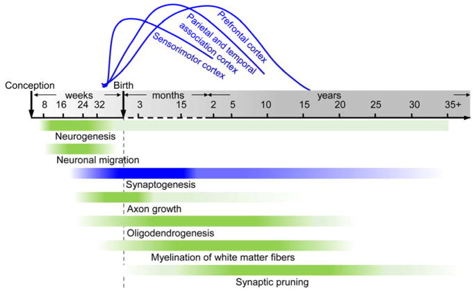
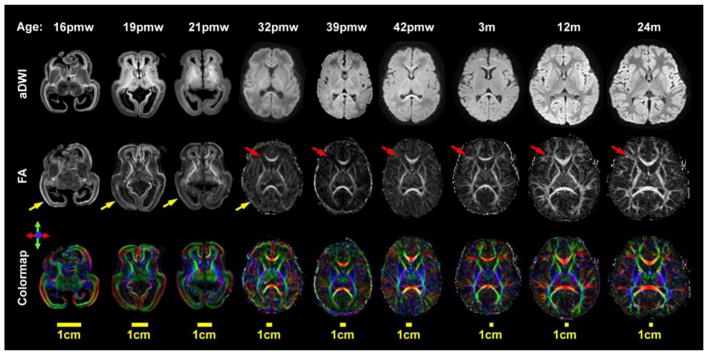
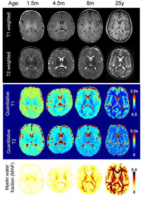
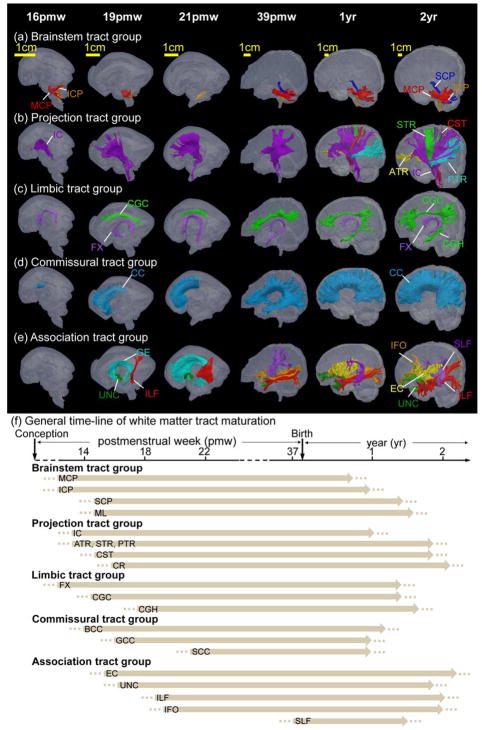
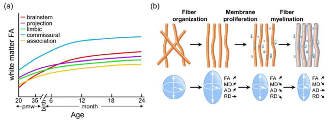
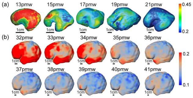
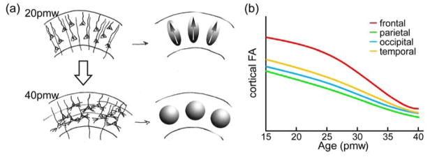
Dynamic macrostructural and microstructural changes take place from the mid-fetal stage to 2 years after birth. Delineating structural changes of the brain during early development provides new insights into the complicated processes of both typical development and the pathological mechanisms underlying various psychiatric and neurological disorders including autism, attention deficit hyperactivity disorder and schizophrenia. Decades of histological studies have identified strong spatial and functional maturation gradients in human brain gray and white matter. The recent improvements in magnetic resonance imaging (MRI) techniques, especially diffusion MRI (dMRI), relaxometry imaging, and magnetization transfer imaging (MTI) have provided unprecedented opportunities to non-invasively quantify and map the early developmental changes at whole brain and regional levels. Here, we review the recent advances in understanding early brain structural development during the second half of gestation and the first two postnatal years using modern MR techniques. Specifically, we review studies that delineate the emergence and microstructural maturation of white matter tracts, as well as dynamic mapping of inhomogeneous cortical microstructural organization unique to fetuses and infants. These imaging studies converge into maturational curves of MRI measurements that are distinctive across different white matter tracts and cortical regions. Furthermore, contemporary models offering biophysical interpretations of the dMRI-derived measurements are illustrated to infer the underlying microstructural changes. Collectively, this review summarizes findings that contribute to charting spatiotemporally heterogeneous gray and white matter structural development, offering MRI-based biomarkers of typical brain development and setting the stage for understanding aberrant brain development in neurodevelopmental disorders.

Keywords: Baby brain; Diffusion MRI; Early development; Microstructure; Quantitative MRI; Tractography.


Figures

Figure 1
Figure 1
Timeline of spatiotemporally distinctive human brain maturational processes, including neurogenesis, neuronal migration, synaptogenesis, axon growth, oligodendrogenesis, myelination of white matter fibers and synaptic pruning. Time axis is in post-conceptional weeks (before birth), postnatal months (until 24 months), and postnatal years (after 2 years). The color intensity in each bar corresponds to the rate of developmental changes. The spatial progression across brain regions is illustrated using synaptogenesis (blue bar) as an example. Specifically, the spatial progression of synaptogenesis from primary sensorimotor cortex to higher-order prefrontal cortex is illustrated by the blue curves above the time axis.
Figure 2
Figure 2
Diffusion MRI (dMRI) contrast in the fetal and infant brain from 16 postmenstrual weeks (pmw) to 2 years (24 months) of age. From top to bottom, axial slices of the averaged diffusion weighted images (aDWI), fractional anisotropy (FA) maps and color-encoded diffusion orientation maps are shown. Yellow arrows indicate high FA values in the cortical plate, and red arrows indicate high FA values in the white matter regions during this early developing period. This figure is generated with the data from Huang lab.
Figure 3
Figure 3
Anatomical images of the developing brain. From top to bottom, T1 weighted, T2 weighted images, quantitative maps of T1, T2 relaxation times (in seconds) and myelin water fraction (MWF) of representative subjects at different ages, infant of 1.5 month (m), 4.5m and 8m and a young adult (25 years), are shown.
Figure 4
Figure 4
Diffusion MRI tractography of the white matter tracts in developing fetal and infant brain from 16 postmenstrual weeks (pmw) to 2 years. (a) Brainstem tracts including inferior, middle, superior cerebellar peduncle (ICP, MCP and SCP) and medial lemniscus (ML); (b) Projection tracts including interior capsule (IC), corona radiata (CR), corticospinal tract (CST), anterior, superior and posterior thalamic radiation (ATR, STR and PTR); (c) Limbic tracts including cingulum bundle in the cingulate cortex (CGC), cingulum bundle in the temporal cortex (CGH) and fornix (FX); (d) Commissural tracts including body, genu and splenium of corpus callosum (BCC, GCC and SCC); (e) Association tracts including fibers in external capsule (EC), inferior longitudinal fasciculus (ILF), inferior occipitofrontal fasciculus (IFO), superior longitudinal fasciculus (SLF) and uncinate fasciculus (UNC). Ganglionic eminence (GE), a transient fetal brain structure and well traced with DTI tractography, is also included in this tract group. (f) General timeline of white matter maturation across different tracts and tract groups. Dotted lines indicate that white matter tracts emerge at these ages, though to a relatively minor degree, and arrows indicate that overall white matter tracts are formed with continuous maturational processes such as myelination and axonal packing thereafter. This figure is generated with the data from Huang lab.
Figure 5
Figure 5
Developmental trajectories of fractional anisotropy (FA) across white matter tracts from the mid-fetal stage to 2 years (a) and the biophysical model of white matter maturation (b). In (a), sketch plots show age-dependent white matter FA changes in five tract groups, namely, brainstem, projection, limbic, commissural and association tract groups from 20 postmenstrual weeks (pmw) to 2 years. Time axis in (a) is in postmenstrual weeks (pmw) before birth and postnatal months until 24 months. In (b), the biophysical model interprets the changes of DTI-derived metrics (i.e. FA, mean/axial/radial diffusivity: MD, AD and RD) (adapted with permission from Dubois et al., 2008 and Qiu et al., 2015).
Figure 6
Figure 6
Mapping of FA onto the cortical surface from 13 to 21 postmenstrual weeks (pmw) in the 2nd trimester (a) (adapted with permission from Huang et al., 2013) and the cortical surface from 32 to 41pmw in the 3rd trimester (b) (adapted with permission from Jeon et al., 2016).
Figure 7
Figure 7
Biophysical model of disruption of radial glial scaffold and associated fractional anisotropy (FA) decrease (a) and distinctive spatiotemporal FA decreases across different cortical regions from 15 to 40 postmenstrual weeks (pmw) (b). In (a), upper left panel demonstrates highly organized radial glial fibers, pyramidal neurons with prominent, radially oriented apical dendrites in a 20pmw brain cortical plate, resulting in the diffusion ellipsoids with high FA values and the primary axes oriented radially (upper right panel); lower left panel demonstrates prominent basal dendrites for the pyramidal cells and thalamocortical afferents disrupting the organized radial organization in a 40pmw brain cortical plate, resulting in the diffusion ellipsoids with low FA values (adapted with permission from McKinstry et al., 2002).

References

    1. Abdelnour F, Voss HU, Raj A. Network diffusion accurately models the relationship between structural and functional brain connectivity networks. Neuroimage. 2014;90:335–347. - PMC - PubMed
    1. Assaf Y, Blumenfeld-Katzir T, Yovel Y, Basser PJ. AxCaliber: a method for measuring axon diameter distribution from diffusion MRI. Magn Reson Med. 2008;59(6):1347–1354. - PMC - PubMed
    1. Atasoy S, Donnelly I, Pearson J. Human brain networks function in connectome-specific harmonic waves. Nat Commun. 2017;7:10340. - PMC - PubMed
    1. Ball G, Srinivasan L, Aljabar P, Counsell SJ, Durighel G, Hajnal JV, Rutherford MA, Edwards AD. Development of cortical microstructure in the preterm human brain. Proc Natl Acad Sci USA. 2013;110(23):9541–9546. - PMC - PubMed
    1. Ball G, Aljabar P, Zebari S, Tusor N, Arichi T, Merchant N, Robinson EC, Ogundipe E, Rueckert D, Edwards AD, Counsell SJ. Rich-club organization of the newborn human brain. Proc Natl Acad Sci USA. 2014;111(20):7456–7461. - PMC - PubMed

Publication types