Review
. 2018 Aug:52:116-123.
doi: 10.1016/j.copbio.2018.03.011. Epub 2018 Apr 12.

Vascularized microfluidic organ-chips for drug screening, disease models and tissue engineering

Affiliations
Review

Vascularized microfluidic organ-chips for drug screening, disease models and tissue engineering

Tatsuya Osaki et al. Curr Opin Biotechnol. 2018 Aug.

Abstract

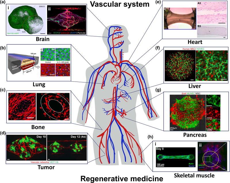
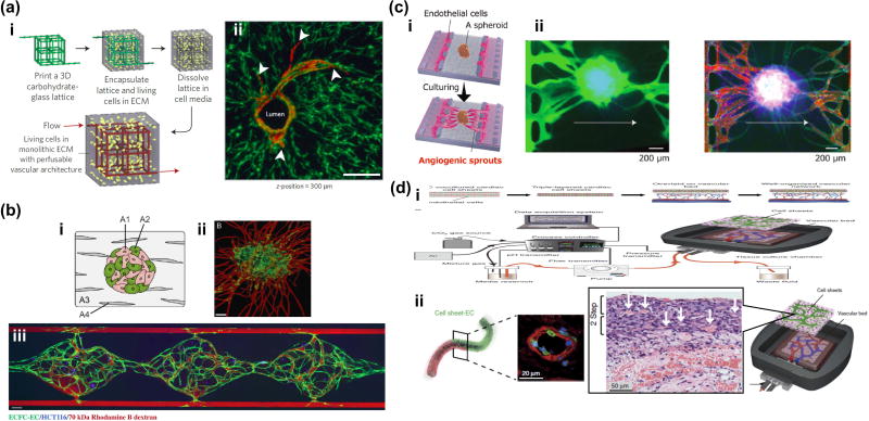
Vascularization of micro-tissues in vitro has enabled formation of tissues larger than those limited by diffusion with appropriate nutrient/gas exchange as well as waste elimination. Furthermore, angiocrine signaling from the vasculature may be essential in mimicking organ-level functions in these micro-tissues. In drug screening applications, the presence of an appropriate blood-organ barrier in the form of a vasculature and its supporting cells (pericytes, appropriate stromal cells) may be essential to reproducing organ-scale drug delivery pharmacokinetics. Cutting-edge techniques including 3D bioprinting and in vitro angiogenesis and vasculogenesis could be applied to vascularize a range of tissues and organoids. Herein, we describe the latest developments in vascularization and prevascularization of micro-tissues and provide an outlook on potential future strategies.


Figures

Figure 1
Figure 1
State-of-the-art of prevascularization and vascularization of micro-tissues in brain (a), lung (b), bone (c), tumor (d), heart (e), liver (f), pancreas (g) and skeletal muscle(h). (a) (i) Fabrication of multi-cellular spheroid by fusing iPS cell-derived human cortical spheroids (hCS) and human subpallium spheroids (hSS) to mimic forebrain-like tissues. (ii) A microfluidic vascularized 3D BBB model developed by co-culturing colony-forming endothelial cells, astrocytes and pericytes in a perfusable microdevice. (b) A microfluidic epithelial and endothelial interface model to simulate the lung alveolar microenvironment. (c) Vascularized bone graft by using 3D bioprinting with biocompatible scaffold. (d) Vascularization of tumor tissues in a perfusable microfluidic platform. (e) Engineering endothelialized hollow-structures in a cardiac muscle fiber in vitro. (f) Prevascularized functional liver bud by spontaneous aggregation of human iPS cell-derived hepatocytes, human umbilical vein endothelial cells (HUVEC) and mesenchymal stem cells (MSC). (g) Prevascularized islet-like tissues with pancreatic β cells, HUVECs, and MSCs. (h) (i) Representative skeletal muscle fiber bundle attached to cantilevers in a microdevice. (ii) Prevascularized skeletal muscle tissue aggregation and anastomosis of prevasculature with host vasculature after transplantation.
Figure 2
Figure 2
Cutting-edge technologies to vascularize tissues in vitro. (a) Engineering of 3D vascular networks in extracellular matrix. (i) 3D casting mold was fabricated with carbohydrate glass lattice by 3D printer. After dissolving the lattice, endothelial cells were seeded into the 3D microchannels. (ii) Endothelialized microchannels and angiogenic sprouting with stromal cells. Potentially, this method could be applied to vascularization of various types of tissues, even relatively large tissues. (b) Vasculogenesis based vascularization in a microfluidic device. Flow of fluorescently tagged dextrans show the perfusability and connectivity of vascular networks to supply sufficient nutrients to HCT116 cells in a gel. This strategy is promising for high-throughput drug screening of vascularized micro-tissues (c) (i) Angiogenesis based vascularization of spheroid in a microfluidic device. (ii) Perfusion of fluorescently tagged dextrans showed that of angiogenic sprouts formed anastomoses with prevascularized networks inside the spheroid, preventing necrotic cell death in the core region. This method could be applied to vascularization of various types of spheroids and organoids for drug testing. (d) Vascularization of sheet-like tissues using an ex vivo vascular bed. Rat vascular bed was extracted and connected to a perfusion system in vitro. Stacked cell sheet with epithelial cells and endothelial cells were placed on this vascular bed. This ex vivo system has promise for the extension of culture time which is currently limited by the lack of vasculature (ii).

References

    1. Nishida K, Yamato M, Hayashida Y, Watanabe K, Yamamoto K, Adachi E, Nagai S, Kikuchi A, Maeda N, Watanabe H, et al. Corneal Reconstruction with Tissue-Engineered Cell Sheets Composed of Autologous Oral Mucosal Epithelium. New England Journal of Medicine. 2004;351:1187–1196. - PubMed
    1. Mandai M, Watanabe A, Kurimoto Y, Hirami Y, Morinaga C, Daimon T, Fujihara M, Akimaru H, Sakai N, Shibata Y, et al. Autologous Induced Stem-Cell–Derived Retinal Cells for Macular Degeneration. New England Journal of Medicine. 2017;376:1038–1046. - PubMed
    1. Atala A, Bauer SB, Soker S, Yoo JJ, Retik AB. Tissue-engineered autologous bladders for patients needing cystoplasty. The Lancet. 2006;367:1241–1246. - PubMed
    1. Memon IA, Sawa Y, Fukushima N, Matsumiya G, Miyagawa S, Taketani S, Sakakida SK, Kondoh H, Aleshin AN, Shimizu T, et al. Repair of impaired myocardium by means of implantation of engineered autologous myoblast sheets. The Journal of Thoracic and Cardiovascular Surgery. 2005;130:1333–1341. - PubMed
    1. Miyagawa S, Domae K, Yoshikawa Y, Fukushima S, Nakamura T, Saito A, Sakata Y, Hamada S, Toda K, Pak K, et al. Phase I Clinical Trial of Autologous Stem Cell–Sheet Transplantation Therapy for Treating Cardiomyopathy. Journal of the American Heart Association. 2017;6 - PMC - PubMed

Publication types