

Shaping eosinophil identity in the tissue contexts of development, homeostasis, and disease
- PMID: 29656559
- PMCID: PMC6013365
- DOI: 10.1002/JLB.1MR1117-442RR
Shaping eosinophil identity in the tissue contexts of development, homeostasis, and disease
Abstract
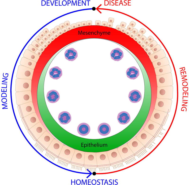
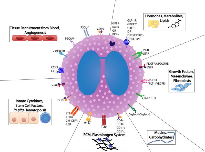
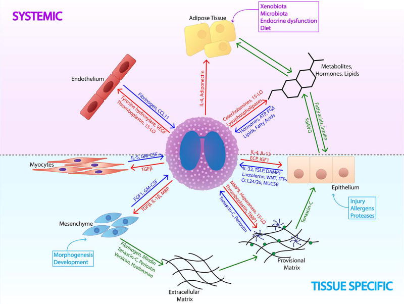
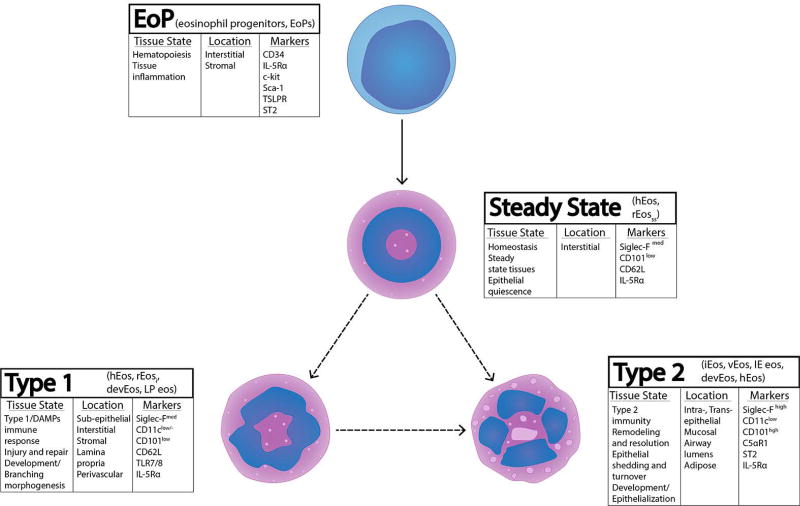
Eosinophils play homeostatic roles in different tissues and are found in several organs at a homeostatic baseline, though their tissue numbers increase significantly in development and disease. The morphological, phenotypical, and functional plasticity of recruited eosinophils are influenced by the dynamic tissue microenvironment changes between homeostatic, morphogenetic, and disease states. Activity of the epithelial-mesenchymal interface, extracellular matrix, hormonal inputs, metabolic state of the environment, as well as epithelial and mesenchymal-derived innate cytokines and growth factors all have the potential to regulate the attraction, retention, in situ hematopoiesis, phenotype, and function of eosinophils. This review examines the reciprocal relationship between eosinophils and such tissue factors, specifically addressing: (1) tissue microenvironments associated with the presence and activity of eosinophils; (2) non-immune tissue ligands regulatory for eosinophil accumulation, hematopoiesis, phenotype, and function (with an emphasis on the extracellular matrix and epithelial-mesenchymal interface); (3) the contribution of eosinophils to regulating tissue biology; (4) eosinophil phenotypic heterogeneity in different tissue microenvironments, classifying eosinophils as progenitors, steady state eosinophils, and Type 1 and 2 activated phenotypes. An appreciation of eosinophil regulation by non-immune tissue factors is necessary for completing the picture of eosinophil immune activation and understanding the functional contribution of these cells to development, homeostasis, and disease.
Keywords: asthma; development; eosinophils; epithelium; extracellular matrix; homeostasis; in situ hematopoiesis; mesenchyme; metabolism; phenotype.
©2018 Society for Leukocyte Biology.
Conflict of interest statement
The authors declare that they have no conflicts of interest.
Figures
References
-
- Mesnil C, Raulier S, Paulissen G, Xiao X, Birrell MA, Pirottin D, Janss T, Starkl P, Ramery E, Henket M, Schleich FN, Radermecker M, Thielemans K, Gillet L, Thiry M, Belvisi MG, Louis R, Desmet C, Marichal T, Bureau F. Lung-resident eosinophils represent a distinct regulatory eosinophil subset. J Clin Invest. 2016;126(9):3279–95. doi: 10.1172/JCI85664. Epub 2016/08/23. - DOI - PMC - PubMed
-
- Jung Y, Wen T, Mingler MK, Caldwell JM, Wang YH, Chaplin DD, Lee EH, Jang MH, Woo SY, Seoh JY, Miyasaka M, Rothenberg ME. IL-1beta in eosinophil-mediated small intestinal homeostasis and IgA production. Mucosal Immunol. 2015;8(4):930–42. doi: 10.1038/mi.2014.123. Epub 2015/01/08. - DOI - PMC - PubMed
-
- de Kleer IM, Kool M, de Bruijn MJ, Willart M, van Moorleghem J, Schuijs MJ, Plantinga M, Beyaert R, Hams E, Fallon PG, Hammad H, Hendriks RW, Lambrecht BN. Perinatal Activation of the Interleukin-33 Pathway Promotes Type 2 Immunity in the Developing Lung. Immunity. 2016;45(6):1285–98. doi: 10.1016/j.immuni.2016.10.031. Epub 2016/12/13. - DOI - PubMed
Publication types
MeSH terms
Substances
Grants and funding
LinkOut - more resources
Full Text Sources
Other Literature Sources
